Chellathurai Amarnath, Ayyamperumal Balaji, Thirumaran Rajakumari, Kathirvelu Gopinathan, Muthaiyan Priya, Kannappan Sivakumar
Department of Radiodiagnosis, Stanley Medical College, Chennai, India.
Department of Radiodiagnosis, Kilpauk Medical College, Chennai, India.
Asian Spine J. 2019 Apr;13(2):189-197. doi: 10.31616/asj.2018.0076. Epub 2018 Nov 27.
Retrospective single institutional observational study.
Segmental spinal dysgenesis (SSD), a complex spinal dysraphic state caused by notochord malformation disorders, is named after its morphological presentation where a spine segment is dysgenetic, malformed or absent. This study's objective was to examine and reassess SSD imaging findings and correlate them with an embryological explanation.
Scott and his colleagues defined SSD as segmental agenesis or dysgenesis of the lumbar or thoracolumbar vertebrae and underlying spinal cord. Tortori-Donati and his colleagues defined it as a morphologic continuum ranging from hypoplasia to an absent spinal cord segment.
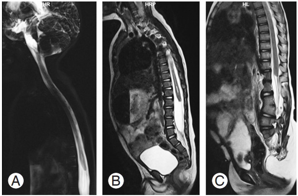
Fifteen children, whose imaging findings and clinical features were consistent with SSD, were included in the study. Magnetic resonance imaging (MRI) was performed per institutional spine protocol.
Five children (33.3%) presented with a high-ending bulbous cord with no caudal segment, six (40%) presented with a dorsal or lumbar segmental dysgenetic cord with a low-lying, bulky caudal cord but without significant spinal canal narrowing, and four (26.6%) presented with segmental caudal dysgenesis with severe kyphoscoliosis, gibbus deformity, and spinal canal narrowing with a normal distal segment (normal or low-lying).
SSD is a complex spinal anomaly in children requiring clinical-radiological assessment followed by multidisciplinary management based on the extent and severity of the dysgenetic cord and the type of SSD. MRI plays a crucial role in both diagnosing and classifying SSD prior to surgical treatment to prevent further impairment.
回顾性单机构观察性研究。
节段性脊柱发育不全(SSD)是一种由脊索畸形障碍引起的复杂脊柱闭合不全状态,因其形态表现而得名,即脊柱节段发育异常、畸形或缺失。本研究的目的是检查和重新评估SSD的影像学表现,并将其与胚胎学解释相关联。
斯科特及其同事将SSD定义为腰椎或胸腰段椎体及下方脊髓的节段性发育不全或发育异常。托尔托里 - 多纳蒂及其同事将其定义为一个从发育不全到脊髓节段缺失的形态连续体。
本研究纳入了15名影像学表现和临床特征与SSD相符的儿童。按照机构脊柱检查方案进行磁共振成像(MRI)检查。
5名儿童(33.3%)表现为高位球茎状脊髓,无尾段;6名(40%)表现为背侧或腰段节段性发育异常脊髓,尾段低位且粗大,但椎管无明显狭窄;4名(26.6%)表现为节段性尾段发育异常,伴有严重脊柱侧凸、驼背畸形以及椎管狭窄,远端节段正常(正常或低位)。
SSD是儿童中的一种复杂脊柱异常,需要进行临床 - 放射学评估,然后根据发育异常脊髓的范围和严重程度以及SSD的类型进行多学科管理。MRI在手术治疗前诊断和分类SSD以防止进一步损伤方面起着关键作用。