García-Peña Claudia M, Ávila-González Daniela, Miquelajáuregui Amaya, Lozano-Flores Carlos, Mastick Grant S, Tamariz Elisa, Varela-Echavarría Alfredo
Department of Developmental Neurobiology and Neurophysiology, Instituto de Neurobiología, Universidad Nacional Autónoma de México (UNAM), Querétaro, México.
Department of Biology, University of Nevada, Reno, Reno, NV, United States.
Front Neuroanat. 2018 Nov 13;12:96. doi: 10.3389/fnana.2018.00096. eCollection 2018.
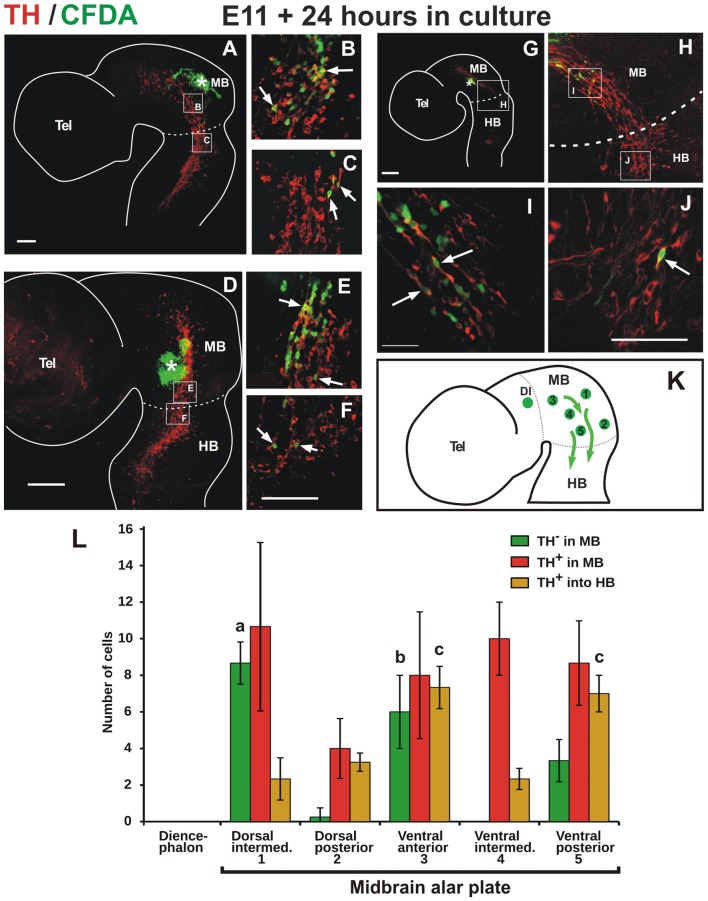
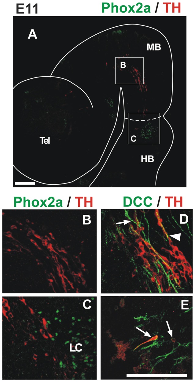
Stereotypic cell migrations in the developing brain are fundamental for the proper patterning of brain regions and formation of neural networks. In this work, we uncovered in the developing rat, a population of neurons expressing tyrosine hydroxylase (TH) that migrates posteriorly from the alar plate of the midbrain, in neurophilic interaction with axons of the mesencephalic nucleus of the trigeminal nerve. A fraction of this population was also shown to traverse the mid-hindbrain boundary, reaching the vicinity of the locus coeruleus (LC) in rhombomere 1 (r1). This migratory population, however, does not have a noradrenergic (NA) phenotype and, in keeping with its midbrain origin, expresses Otx2 which is down regulated upon migration into the hindbrain. The interaction with the trigeminal mesencephalic axons is necessary for the arrangement and distribution of migratory cells as these aspects are dramatically altered in whole embryo cultures upon disruption of trigeminal axon projection by interfering with DCC function. Moreover, in mouse embryos in an equivalent developmental stage, we detected a cell population that also migrates caudally within the midbrain apposed to mesencephalic trigeminal axons but that does not express TH; a fraction of this population expresses calbindin instead. Overall, our work identified TH-expressing neurons from the rat midbrain alar plate that migrate tangentially over long distances within the midbrain and into the hindbrain by means of a close interaction with trigeminal mesencephalic axons. A different migratory population in this region and also in mouse embryos revealed diversity among the cells that follow this descending migratory pathway.
在发育中的大脑中,刻板的细胞迁移对于脑区的正确模式形成和神经网络的形成至关重要。在这项研究中,我们在发育中的大鼠中发现了一群表达酪氨酸羟化酶(TH)的神经元,它们从中脑翼板向后迁移,与三叉神经中脑核的轴突发生嗜神经相互作用。这一群体中的一部分还被证明穿过中后脑边界,到达菱脑节1(r1)中蓝斑(LC)附近。然而,这个迁移群体不具有去甲肾上腺素能(NA)表型,并且与其中脑起源一致,表达Otx2,而在迁移到后脑时该基因表达下调。与三叉神经中脑轴突的相互作用对于迁移细胞的排列和分布是必要的,因为在全胚胎培养中,当通过干扰DCC功能破坏三叉神经轴突投射时,这些方面会发生显著改变。此外,在处于等效发育阶段的小鼠胚胎中,我们检测到一群细胞也在中脑内沿着与三叉神经中脑轴突相邻的方向向尾侧迁移,但不表达TH;这一群体中的一部分转而表达钙结合蛋白。总体而言,我们的研究确定了来自大鼠中脑翼板的表达TH的神经元,它们通过与三叉神经中脑轴突的紧密相互作用,在中脑内并向后脑进行长距离的切向迁移。在该区域以及小鼠胚胎中的另一群迁移细胞揭示了沿着这条下行迁移途径的细胞之间的多样性。