Yin Zhiyang, Chang Miao, Wei Shengnan, Jiang Xiaowei, Zhou Yifang, Cui Lingling, Lv Jing, Wang Fei, Tang Yanqing
Department of Psychiatry, The First Affiliated Hospital of China Medical University, Shenyang, China.
Brain Function Research Section, The First Affiliated Hospital of China Medical University, Shenyang, China.
Front Neurosci. 2018 Nov 14;12:842. doi: 10.3389/fnins.2018.00842. eCollection 2018.
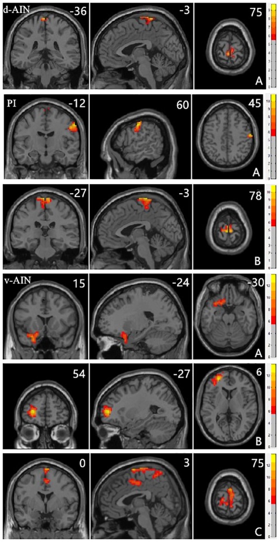
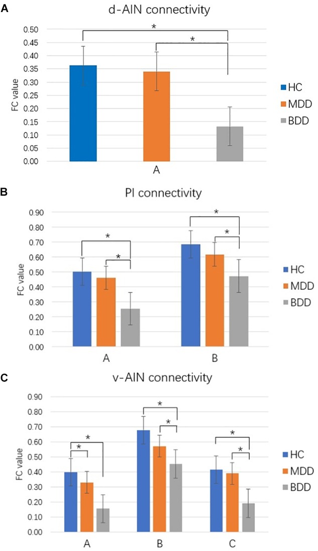
Clinically, it is very difficult to distinguish between major depressive disorder (MDD) and bipolar disorder (BD) in the period of depression. Increasing evidence shows that the insula plays an important role in depression. We aimed to compare the resting-state functional connectivity (rsFC) of insular subregions in patients with MDD and BD in depressive episodes (BDD), who had never experienced manic or hypomanic episodes when they were scanned to identify biomarkers for the identification of two diseases. We recruited 21 BDD patients, 40 MDD patients and 70 healthy controls (HC). Resting-state functional magnetic resonance imaging (rs-fMRI) was performed. BDD patients had never had manic or hypomanic episodes when they were scanned, and the diagnoses were determined by follow-up. We divided the insula into three parts including the ventral anterior insular cortex (v-AIN), dorsal anterior insular cortex (d-AIN), and posterior insula (PI). The insular-based rsFC was compared among the three groups, and an analysis of the correlation between the rsFC value and Hamilton depression and anxiety scales was carried out. BDD and MDD patients demonstrated decreased rsFC from the v-AIN to the left superior/middle frontal gyrus compared with the HC group. Versus MDD and HC groups, BDD patients exhibited decreased rsFC from the v-AIN to the area in the left orbital frontal gyrus and left superior temporal gyrus (included temporal pole), from the PI to the right lateral postcentral gyrus and from all three insular subregions to the somatosensory and motor cortex. Meanwhile, a correlation between the rsFC value of the PI-right lateral postcentral gyrus and anxiety score was observed in patients. Our findings show BDD and MDD patients have similar decreases in insular connectivity in the dorsal lateral frontal regions, and BDD patients have specific decreased insular connectivity, especially in the somatosensory and motor cortex, which may be used as imaging evidence for clinical identification.
临床上,在抑郁期很难区分重度抑郁症(MDD)和双相情感障碍(BD)。越来越多的证据表明,脑岛在抑郁症中起重要作用。我们旨在比较MDD患者和BD抑郁发作期(BDD)患者脑岛亚区域的静息态功能连接(rsFC),这些患者在扫描时从未经历过躁狂或轻躁狂发作,以识别用于鉴别这两种疾病的生物标志物。我们招募了21名BDD患者、40名MDD患者和70名健康对照者(HC)。进行了静息态功能磁共振成像(rs-fMRI)。BDD患者在扫描时从未有过躁狂或轻躁狂发作,诊断通过随访确定。我们将脑岛分为三个部分,包括腹侧前脑岛皮质(v-AIN)、背侧前脑岛皮质(d-AIN)和后脑岛(PI)。比较了三组之间基于脑岛的rsFC,并对rsFC值与汉密尔顿抑郁和焦虑量表之间的相关性进行了分析。与HC组相比,BDD和MDD患者从v-AIN到左侧额上/中回的rsFC降低。与MDD和HC组相比,BDD患者从v-AIN到左侧眶额回和左侧颞上回(包括颞极)区域、从PI到右侧中央后回以及从所有三个脑岛亚区域到体感和运动皮层的rsFC降低。同时,在患者中观察到PI-右侧中央后回的rsFC值与焦虑评分之间存在相关性。我们的研究结果表明,BDD和MDD患者在背外侧额叶区域的脑岛连接性有相似的降低,并且BDD患者有特定的脑岛连接性降低,特别是在体感和运动皮层,这可能作为临床鉴别的影像学证据。