Department of Physiology, Development and Neuroscience, University of Cambridge, Downing Street, Cambridge CB2 3DY, UK.
Department of Physiology, Development and Neuroscience, University of Cambridge, Downing Street, Cambridge CB2 3DY, UK.
Dev Cell. 2018 Dec 17;47(6):727-740.e6. doi: 10.1016/j.devcel.2018.10.029. Epub 2018 Nov 29.
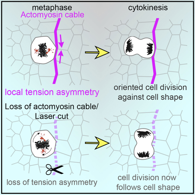
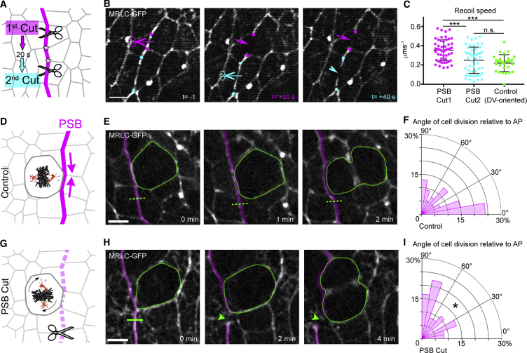
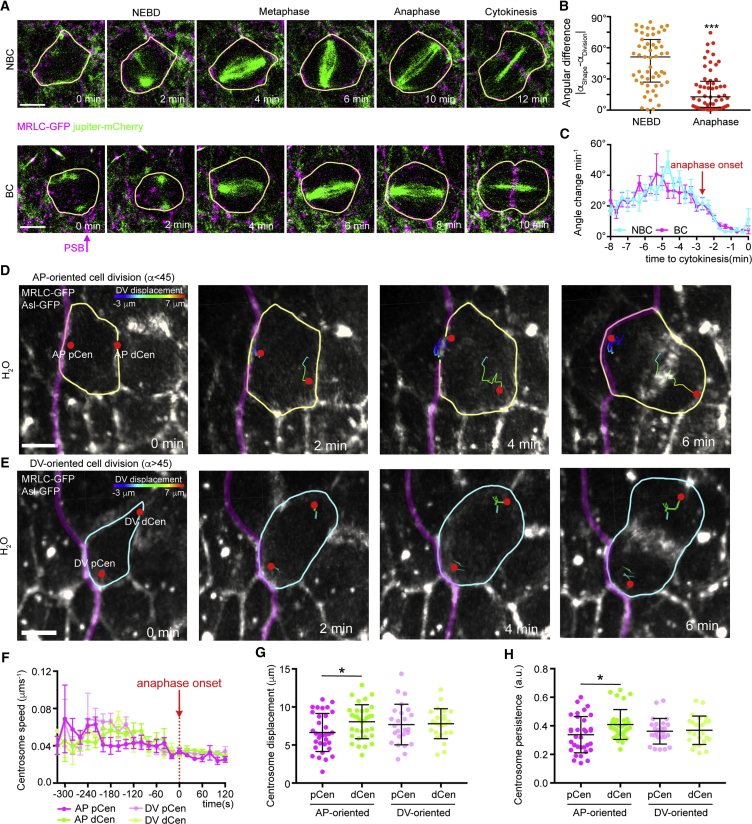
Cell shape is known to influence the plane of cell division. In vitro, mechanical constraints can also orient mitoses; however, in vivo it is not clear whether tension can orient the mitotic spindle directly, because tissue-scale forces can change cell shape. During segmentation of the Drosophila embryo, actomyosin is enriched along compartment boundaries forming supracellular cables that keep cells segregated into distinct compartments. Here, we show that these actomyosin cables orient the planar division of boundary cells perpendicular to the boundaries. This bias overrides the influence of cell shape, when cells are mildly elongated. By decreasing actomyosin cable tension with laser ablation or, conversely, ectopically increasing tension with laser wounding, we demonstrate that local tension is necessary and sufficient to orient mitoses in vivo. This involves capture of the spindle pole by the actomyosin cortex. These findings highlight the importance of actomyosin-mediated tension in spindle orientation in vivo.
细胞形状已知会影响细胞分裂的平面。在体外,机械约束也可以定向有丝分裂;然而,在体内尚不清楚张力是否可以直接定向有丝分裂纺锤体,因为组织尺度的力可以改变细胞形状。在果蝇胚胎的分割过程中,肌动球蛋白沿隔室边界富集,形成超细胞电缆,使细胞分隔成不同的隔室。在这里,我们表明这些肌动球蛋白电缆将边界细胞的平面分裂垂直于边界定向。当细胞轻度伸长时,这种偏差会覆盖细胞形状的影响。通过激光消融降低肌动球蛋白电缆张力,或者相反地,通过激光损伤异位增加张力,我们证明局部张力是体内定向有丝分裂所必需和充分的。这涉及到纺锤体极被肌动球蛋白皮质捕获。这些发现强调了肌动球蛋白介导的张力在体内纺锤体定向中的重要性。