School of Life Sciences, University of Science and Technology of China, Hefei, Anhui, P.R. China.
Department of Microbiology, Immunology & Molecular Genetics, University of California, Los Angeles (UCLA), Los Angeles, California, United States of America.
PLoS Pathog. 2018 Dec 3;14(12):e1007452. doi: 10.1371/journal.ppat.1007452. eCollection 2018 Dec.
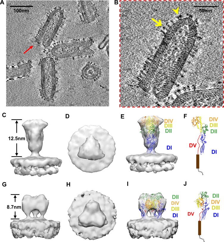
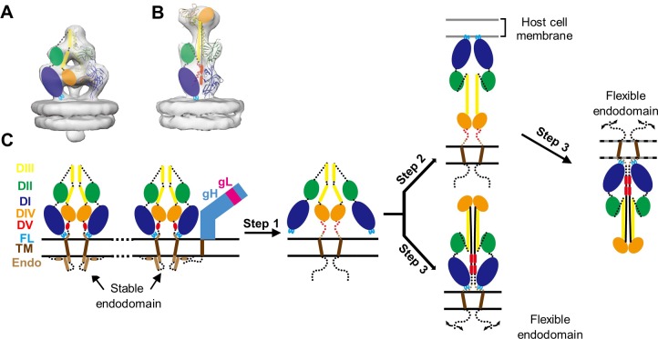
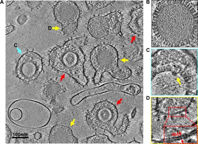
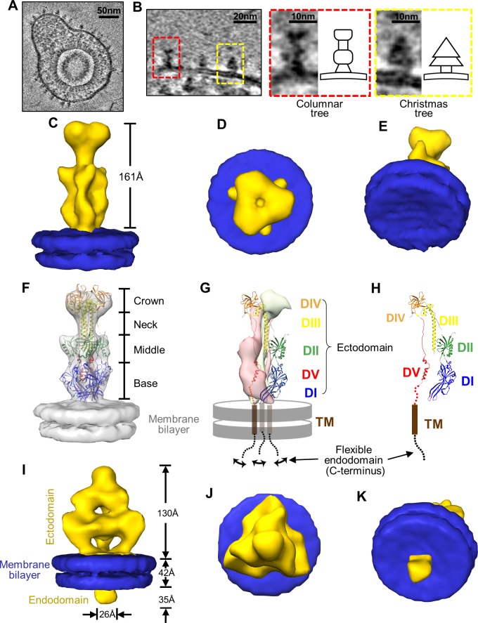
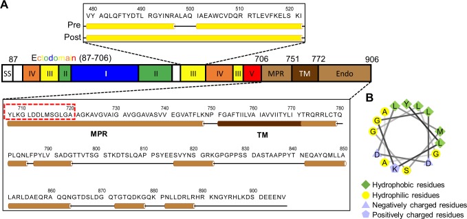
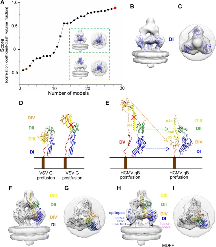
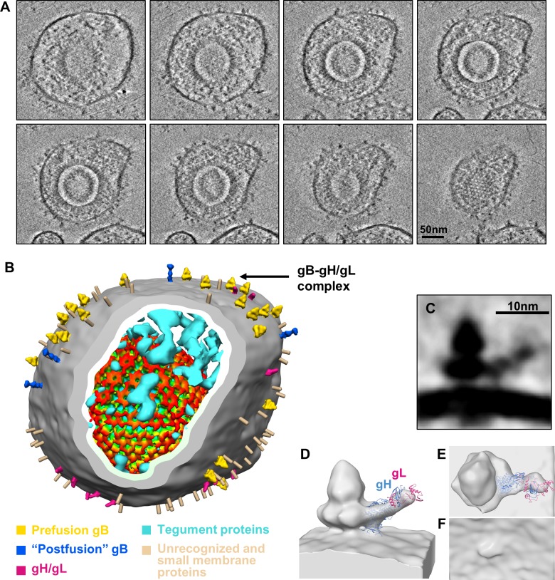
Human cytomegalovirus (HCMV) enters host by glycoprotein B (gB)-mediated membrane fusion upon receptor-binding to gH/gL-related complexes, causing devastating diseases such as birth defects. Although an X-ray crystal structure of the recombinant gB ectodomain at postfusion conformation is available, the structures of prefusion gB and its complex with gH/gL on the viral envelope remain elusive. Here, we demonstrate the utility of cryo electron tomography (cryoET) with energy filtering and the cutting-edge technologies of Volta phase plate (VPP) and direct electron-counting detection to capture metastable prefusion viral fusion proteins and report the structures of glycoproteins in the native environment of HCMV virions. We established the validity of our approach by obtaining cryoET in situ structures of the vesicular stomatitis virus (VSV) glycoprotein G trimer (171 kD) in prefusion and postfusion conformations, which agree with the known crystal structures of purified G trimers in both conformations. The excellent contrast afforded by these technologies has enabled us to identify gB trimers (303kD) in two distinct conformations in HCMV tomograms and obtain their in situ structures at up to 21 Å resolution through subtomographic averaging. The predominant conformation (79%), which we designate as gB prefusion conformation, fashions a globular endodomain and a Christmas tree-shaped ectodomain, while the minority conformation (21%) has a columnar tree-shaped ectodomain that matches the crystal structure of the "postfusion" gB ectodomain. We also observed prefusion gB in complex with an "L"-shaped density attributed to the gH/gL complex. Integration of these structures of HCMV glycoproteins in multiple functional states and oligomeric forms with existing biochemical data and domain organization of other class III viral fusion proteins suggests that gH/gL receptor-binding triggers conformational changes of gB endodomain, which in turn triggers two essential steps to actuate virus-cell membrane fusion: exposure of gB fusion loops and unfurling of gB ectodomain.
人类巨细胞病毒 (HCMV) 通过糖蛋白 B (gB) 介导的膜融合进入宿主,在与 gH/gL 相关复合物结合后受体,导致毁灭性疾病,如出生缺陷。尽管已有融合后构象的重组 gB 外域的 X 射线晶体结构,但病毒包膜上的 gB 前融合构象及其与 gH/gL 的复合物的结构仍然难以捉摸。在这里,我们展示了低温电子断层扫描 (cryoET) 与能量过滤以及尖端技术(如 Volta 相板 (VPP) 和直接电子计数检测)的结合使用的效用,以捕获不稳定的前融合病毒融合蛋白,并报告 HCMV 病毒粒子的天然环境中的糖蛋白结构。我们通过获得水疱性口炎病毒 (VSV) 糖蛋白 G 三聚体 (171 kD) 的融合前和融合后构象的 cryoET 原位结构,证明了我们方法的有效性,这些结构与两种构象中已知的纯化 G 三聚体的晶体结构一致。这些技术提供的优异对比度使我们能够在 HCMV 断层图像中识别出两种不同构象的 gB 三聚体(303kD),并通过亚断层平均获得高达 21 Å 的分辨率的原位结构。我们将主要构象(79%)指定为 gB 前融合构象,其塑造了一个球形的内域和一个圣诞树形状的外域,而少数构象(21%)具有柱状树状的外域,与“融合后”gB 外域的晶体结构相匹配。我们还观察到与 gH/gL 复合物相关的“L”形密度的前融合 gB。将这些多种功能状态和寡聚形式的 HCMV 糖蛋白结构与现有生化数据和其他 III 类病毒融合蛋白的结构域组织整合在一起,表明 gH/gL 受体结合触发 gB 内域的构象变化,这反过来又触发了激活病毒-细胞膜融合的两个基本步骤:gB 融合环的暴露和 gB 外域的展开。