• 文献检索
  • 文档翻译
  • 深度研究
  • 学术资讯
  • Suppr Zotero 插件Zotero 插件
  • 邀请有礼
  • 套餐&价格
  • 历史记录
应用&插件
Suppr Zotero 插件Zotero 插件浏览器插件Mac 客户端Windows 客户端微信小程序
定价
高级版会员购买积分包购买API积分包
服务
文献检索文档翻译深度研究API 文档MCP 服务
关于我们
关于 Suppr公司介绍联系我们用户协议隐私条款
关注我们

Suppr 超能文献

核心技术专利:CN118964589B侵权必究
粤ICP备2023148730 号-1Suppr @ 2026

文献检索

告别复杂PubMed语法,用中文像聊天一样搜索,搜遍4000万医学文献。AI智能推荐,让科研检索更轻松。

立即免费搜索

文件翻译

保留排版,准确专业,支持PDF/Word/PPT等文件格式,支持 12+语言互译。

免费翻译文档

深度研究

AI帮你快速写综述,25分钟生成高质量综述,智能提取关键信息,辅助科研写作。

立即免费体验

K3.1 蛋白在大鼠左心室游离壁呈跨壁梯度表达。

K 3.1 protein is expressed as a transmural gradient across the rat left ventricular free wall.

机构信息

Department of Biomedical Sciences, Department Faculty of Health Sciences, University of Hull, Hull, UK.

Centre de Recherche Cardio-Thoracique de Bordeaux U1045, Université de Bordeaux, Bordeaux, France.

出版信息

J Cardiovasc Electrophysiol. 2019 Mar;30(3):383-391. doi: 10.1111/jce.13805. Epub 2018 Dec 28.

DOI:10.1111/jce.13805
PMID:30516300
原文链接:https://pmc.ncbi.nlm.nih.gov/articles/PMC6446730/
Abstract

INTRODUCTION

K 3.1, also known as TASK-1, is a twin-pore acid-sensitive repolarizing K channel, responsible for a background potassium current that significantly contributes to setting the resting membrane potential of cardiac myocytes. Inhibition of I alters cardiac repolarization and is proarrhythmogenic. In this study, we have examined the expression of K 3.1 and function of this channel in tissue and myocytes from across the left ventricular free wall.

METHODS AND RESULTS

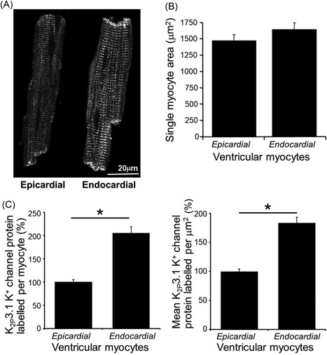
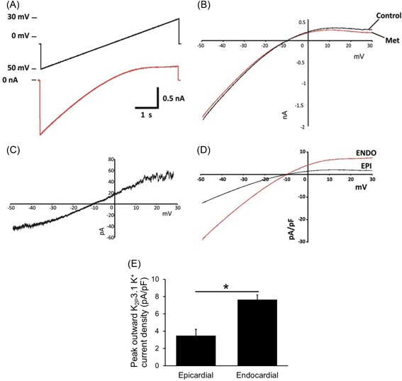
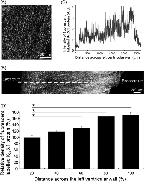
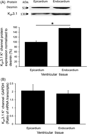
Using fluorescence immunocytochemistry, the expression of K 3.1 protein in myocytes from the subendocardial region was found to be twice (205% ± 13.5%) that found in myocytes from the subepicardial region of the left ventricle (100% ± 5.3%). The left ventricular free wall exhibited a marked transmural gradient of K 3.1 protein expression. Western blot analysis confirmed significantly higher K 3.1 protein expression in subendocardial tissue (156% ± 2.5%) than subepicardial tissue (100% ± 5.0%). However, there was no difference in K 3.1 messenger RNA expression. Whole-cell patch clamp identified I current density to be significantly greater in myocytes isolated from the subendocardium (7.66 ± 0.53 pA/pF) compared with those from the subepicardium (3.47 ± 0.74 pA/pF).

CONCLUSIONS

This is the first study to identify a transmural gradient of K 3.1 in the left ventricle. This gradient has implications for understanding ventricular arrhythmogenesis under conditions of ischemia but also in response to other modulatory factors, such as adrenergic stimulation and the presence of anesthetics that inhibits or activates this channel.

摘要

简介

K 3.1,也称为 TASK-1,是一种双孔酸敏感复极化钾通道,负责背景钾电流,对心肌细胞的静息膜电位有重要贡献。I 的抑制会改变心脏复极化,导致心律失常。在这项研究中,我们检查了左心室游离壁各处组织和心肌细胞中 K 3.1 的表达和功能。

方法和结果

使用荧光免疫细胞化学,发现心内膜下区域心肌细胞中 K 3.1 蛋白的表达是心外膜下区域心肌细胞(100%±5.3%)的两倍(205%±13.5%)。左心室游离壁表现出明显的 K 3.1 蛋白表达的穿壁梯度。Western blot 分析证实心内膜下组织中 K 3.1 蛋白表达明显高于心外膜下组织(156%±2.5%比 100%±5.0%)。然而,K 3.1 信使 RNA 表达没有差异。全细胞膜片钳技术鉴定出心内膜下分离的心肌细胞 I 电流密度(7.66±0.53 pA/pF)明显大于心外膜下分离的心肌细胞(3.47±0.74 pA/pF)。

结论

这是第一项研究表明左心室存在 K 3.1 的穿壁梯度。这种梯度对于理解缺血条件下的心室心律失常发生机制具有重要意义,但也对其他调节因素(如肾上腺素能刺激和抑制或激活这种通道的麻醉剂)具有重要意义。

https://cdn.ncbi.nlm.nih.gov/pmc/blobs/76d9/6446730/3ae287aa8cb0/JCE-30-383-g004.jpg
https://cdn.ncbi.nlm.nih.gov/pmc/blobs/76d9/6446730/448347ef380c/JCE-30-383-g001.jpg
https://cdn.ncbi.nlm.nih.gov/pmc/blobs/76d9/6446730/a784207670a7/JCE-30-383-g002.jpg
https://cdn.ncbi.nlm.nih.gov/pmc/blobs/76d9/6446730/93a6ac290ddb/JCE-30-383-g003.jpg
https://cdn.ncbi.nlm.nih.gov/pmc/blobs/76d9/6446730/3ae287aa8cb0/JCE-30-383-g004.jpg

相似文献

1
K 3.1 protein is expressed as a transmural gradient across the rat left ventricular free wall.K3.1 蛋白在大鼠左心室游离壁呈跨壁梯度表达。
J Cardiovasc Electrophysiol. 2019 Mar;30(3):383-391. doi: 10.1111/jce.13805. Epub 2018 Dec 28.
2
Atrial fibrillation and heart failure-associated remodeling of two-pore-domain potassium (K) channels in murine disease models: focus on TASK-1.心房颤动和心力衰竭相关的双孔钾 (K) 通道在小鼠疾病模型中的重构:聚焦 TASK-1。
Basic Res Cardiol. 2018 Jun 7;113(4):27. doi: 10.1007/s00395-018-0687-9.
3
Contribution of two-pore K channels to cardiac ventricular action potential revealed using human iPSC-derived cardiomyocytes.利用人诱导多能干细胞衍生的心肌细胞揭示双孔钾通道对心脏心室动作电位的作用。
Am J Physiol Heart Circ Physiol. 2017 Jun 1;312(6):H1144-H1153. doi: 10.1152/ajpheart.00107.2017. Epub 2017 Mar 24.
4
pH-dependent inhibition of K₂P3.1 prolongs atrial refractoriness in whole hearts.pH依赖性对K₂P3.1的抑制作用可延长完整心脏的心房不应期。
Pflugers Arch. 2016 Apr;468(4):643-54. doi: 10.1007/s00424-015-1779-0. Epub 2016 Jan 5.
5
Repolarizing K+ currents in nonfailing human hearts. Similarities between right septal subendocardial and left subepicardial ventricular myocytes.正常人类心脏中的复极化钾电流。右间隔心内膜下和左心室外膜下心室肌细胞之间的相似性。
Circulation. 1995 Sep 1;92(5):1179-87. doi: 10.1161/01.cir.92.5.1179.
6
The effects of halothane, isoflurane, and sevoflurane on Ca2+ current and transient outward K+ current in subendocardial and subepicardial myocytes from the rat left ventricle.氟烷、异氟烷和七氟烷对大鼠左心室心内膜下和心外膜下心肌细胞Ca2+电流和瞬时外向K+电流的影响。
Anesth Analg. 2004 Dec;99(6):1615-1622. doi: 10.1213/01.ANE.0000138422.40560.A9.
7
Acid-sensitive TWIK and TASK two-pore domain potassium channels change ion selectivity and become permeable to sodium in extracellular acidification.酸敏感的 TWIK 和 TASK 双孔钾通道在细胞外酸化时改变离子选择性并对钠离子通透。
J Biol Chem. 2012 Oct 26;287(44):37145-53. doi: 10.1074/jbc.M112.398164. Epub 2012 Sep 4.
8
Gene expression of stretch-activated channels and mechanoelectric feedback in the heart.心脏中牵张激活通道的基因表达与机械电反馈
Clin Exp Pharmacol Physiol. 2006 Jul;33(7):642-8. doi: 10.1111/j.1440-1681.2006.04392.x.
9
A study on the electrophysiological heterogeneity of rabbit ventricular myocytes the effect of ischemia on action potentials and potassium currents.一项关于兔心室肌细胞电生理异质性:缺血对动作电位和钾电流影响的研究。
Sheng Li Xue Bao. 2000 Oct;52(5):360-4.
10
Evidence for cystic fibrosis transmembrane conductance regulator chloride current in swine ventricular myocytes.猪心室肌细胞中囊性纤维化跨膜传导调节因子氯离子电流的证据。
J Mol Cell Cardiol. 2007 Jan;42(1):98-105. doi: 10.1016/j.yjmcc.2006.10.002. Epub 2006 Nov 16.

本文引用的文献

1
TASK-1 current is inhibited by phosphorylation during human and canine chronic atrial fibrillation.在人类和犬类慢性心房颤动期间,TASK-1电流受到磷酸化的抑制。
Am J Physiol Heart Circ Physiol. 2015 Jan 15;308(2):H126-34. doi: 10.1152/ajpheart.00614.2014. Epub 2014 Nov 26.
2
Cloning, functional characterization, and remodeling of K2P3.1 (TASK-1) potassium channels in a porcine model of atrial fibrillation and heart failure.心房颤动和心力衰竭猪模型中K2P3.1(TASK-1)钾通道的克隆、功能表征及重塑
Heart Rhythm. 2014 Oct;11(10):1798-805. doi: 10.1016/j.hrthm.2014.06.020. Epub 2014 Jun 18.
3
The inhibition of the potassium channel TASK-1 in rat cardiac muscle by endothelin-1 is mediated by phospholipase C.
内皮素-1 通过磷脂酶 C 抑制大鼠心肌钾通道 TASK-1。
Cardiovasc Res. 2013 Jan 1;97(1):97-105. doi: 10.1093/cvr/cvs285. Epub 2012 Sep 12.
4
TASK-1 channels may modulate action potential duration of human atrial cardiomyocytes.TASK-1通道可能调节人心房心肌细胞的动作电位持续时间。
Cell Physiol Biochem. 2011;28(4):613-24. doi: 10.1159/000335757. Epub 2011 Dec 14.
5
Knock-out of the potassium channel TASK-1 leads to a prolonged QT interval and a disturbed QRS complex.钾通道TASK-1的敲除导致QT间期延长和QRS波群紊乱。
Cell Physiol Biochem. 2011;28(1):77-86. doi: 10.1159/000331715. Epub 2011 Aug 16.
6
Functional role of TASK-1 in the heart: studies in TASK-1-deficient mice show prolonged cardiac repolarization and reduced heart rate variability.TASK-1 在心脏中的功能作用:在 TASK-1 缺陷型小鼠中的研究表明心脏复极化延长和心率变异性降低。
Basic Res Cardiol. 2011 Jan;106(1):75-87. doi: 10.1007/s00395-010-0128-x. Epub 2010 Oct 27.
7
The human cardiac K2P3.1 (TASK-1) potassium leak channel is a molecular target for the class III antiarrhythmic drug amiodarone.人类心脏 K2P3.1(TASK-1)钾泄漏通道是 III 类抗心律失常药物胺碘酮的分子靶点。
Naunyn Schmiedebergs Arch Pharmacol. 2010 Mar;381(3):261-70. doi: 10.1007/s00210-009-0454-4. Epub 2009 Sep 24.
8
Differential effects of volatile and intravenous anesthetics on the activity of human TASK-1.挥发性和静脉麻醉剂对人TASK-1活性的不同影响。
Am J Physiol Cell Physiol. 2007 Oct;293(4):C1319-26. doi: 10.1152/ajpcell.00100.2007. Epub 2007 Aug 15.
9
The acid-sensitive potassium channel TASK-1 in rat cardiac muscle.大鼠心肌中的酸敏感钾通道TASK-1
Cardiovasc Res. 2007 Jul 1;75(1):59-68. doi: 10.1016/j.cardiores.2007.02.025. Epub 2007 Feb 28.
10
Molecular physiology of cardiac repolarization.心脏复极化的分子生理学
Physiol Rev. 2005 Oct;85(4):1205-53. doi: 10.1152/physrev.00002.2005.