Hamid Mohamed A, Mehta Mitul C, Kuppermann Baruch D
Gavin Herbert Eye Institute, University of California Irvine, 850 Health Sciences Road, Irvine, CA 92697 USA.
Int J Retina Vitreous. 2018 Nov 30;4:45. doi: 10.1186/s40942-018-0147-6. eCollection 2018.
Prader-Willi syndrome (PWS) is a genetic disease caused by loss of expression of the paternally inherited copy of several genes on the long arm of chromosome 15. Ophthalmic manifestations of PWS include strabismus, amblyopia, nystagmus, hypopigmentation of the iris and choroid, diabetic retinopathy, cataract and congenital ectropion uvea. An overlap between PWS and oculocutaneous albinism (OCA) has long been recognized and attributed to deletion of OCA2 gene located in PWS critical region (PWCR).
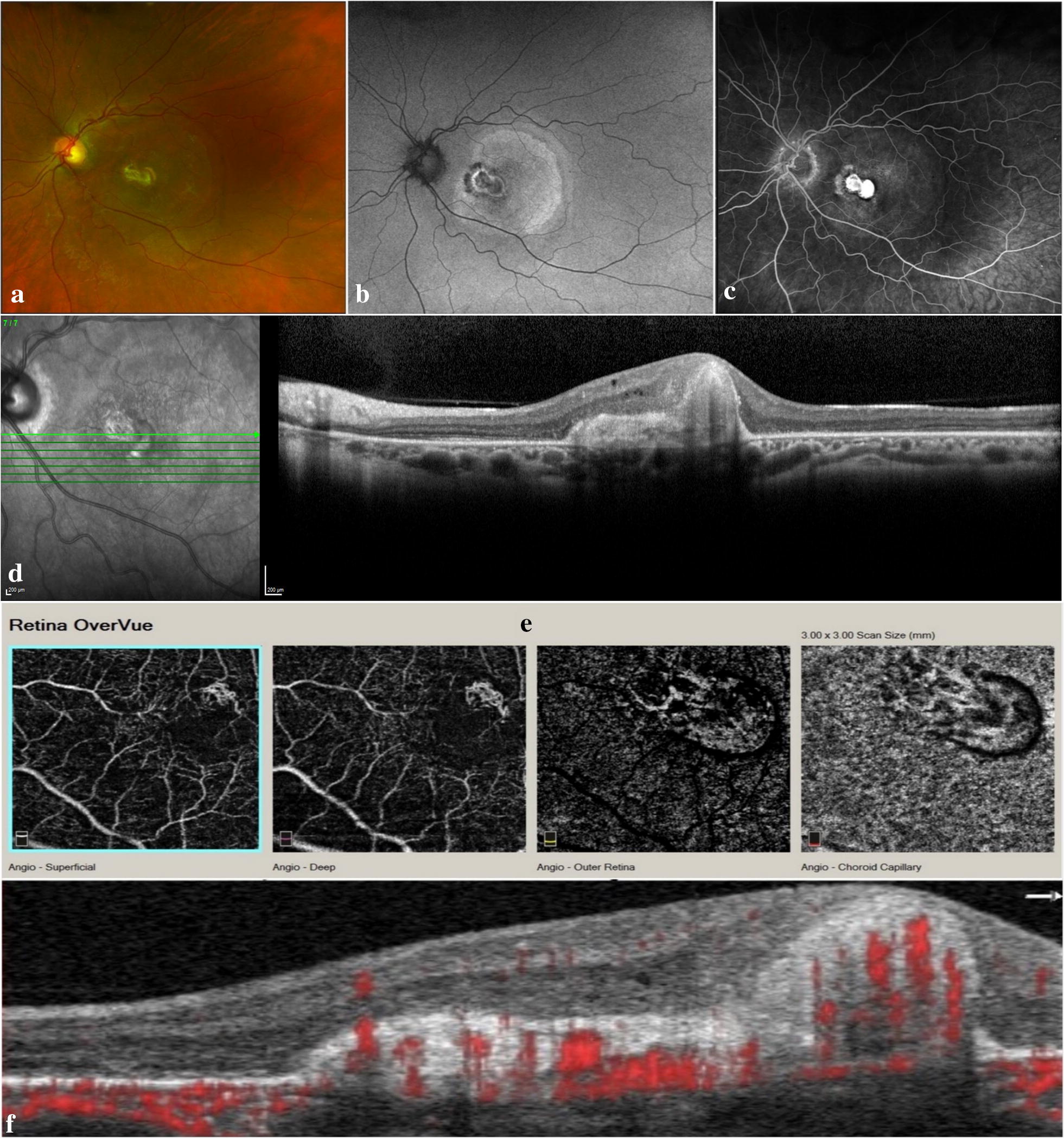
A 30-year-old male patient with PWS presented with vision loss in his left eye. His right eye had normal visual acuity. Multimodal imaging revealed absence of a foveal depression and extremely reduced diameter of the foveal avascular zone in the right eye and an inactive type 2 macular neovascular lesion in the left eye.
We report a presumed association of fovea plana and choroidal neovascularization with PWS. The use of multimodal imaging revealed novel findings in a PWS patient that might enrich our current understanding of the overlap between PWS and OCA.
普拉德-威利综合征(PWS)是一种由15号染色体长臂上若干父系遗传基因拷贝的表达缺失引起的遗传性疾病。PWS的眼部表现包括斜视、弱视、眼球震颤、虹膜和脉络膜色素减退、糖尿病性视网膜病变、白内障和先天性葡萄膜外翻。PWS与眼皮肤白化病(OCA)之间的重叠早已被认识到,并归因于位于PWS关键区域(PWCR)的OCA2基因缺失。
一名30岁的PWS男性患者左眼视力丧失。他的右眼视力正常。多模态成像显示右眼无中央凹凹陷且中央凹无血管区直径极度减小,左眼有非活动性2型黄斑新生血管病变。
我们报告了扁平中央凹和脉络膜新生血管与PWS之间的推测关联。多模态成像的应用在一名PWS患者中发现了新的发现,这可能丰富我们目前对PWS与OCA重叠的理解。