Heart Hospital, Tampere University Hospital and Faculty of Medicine and Life Sciences, University of Tampere, Tampere, Finland.
Division of Image Processing, Department of Radiology, Leiden University Medical Center, Leiden, Netherlands.
PLoS One. 2018 Dec 17;13(12):e0209110. doi: 10.1371/journal.pone.0209110. eCollection 2018.
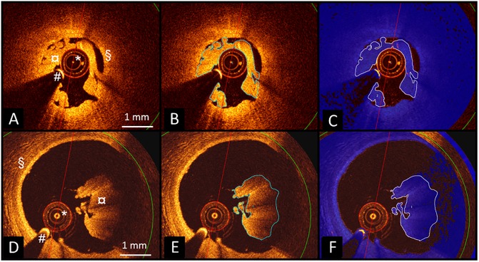
Analysis of intracoronary thrombus type by optical coherence tomography (OCT) imaging is highly subjective. We aimed to compare a newly developed image analysis method to subjective visual classification of thrombus type identified by OCT.
Thirty patients with acute ST elevation myocardial infarction were included. Thrombus type visually classified by two independent readers was compared with analysis using QCU-CMS software.
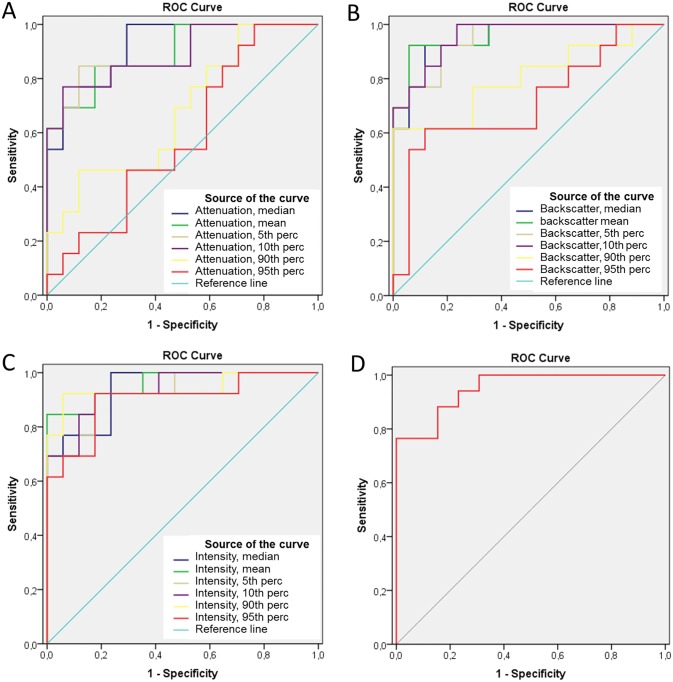
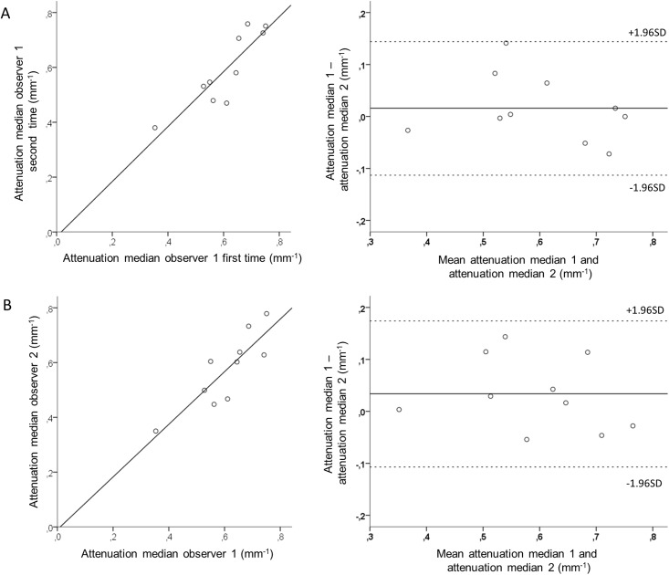
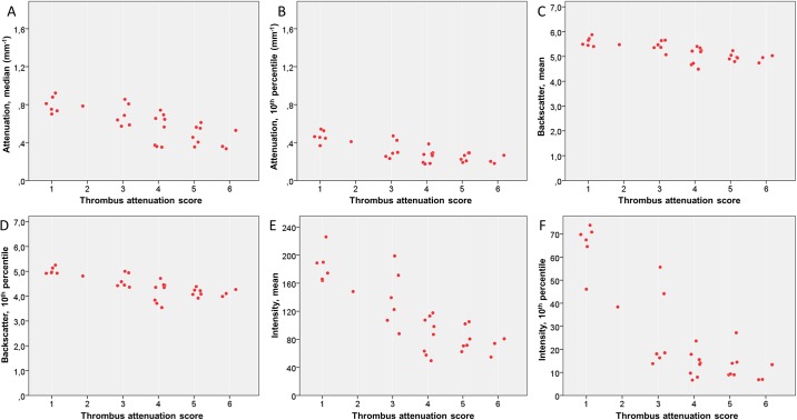
Repeatability of the computer-based measurements was good. By using a ROC, area under curve values for discrimination of white and red thrombi were 0.92 (95% confidence intervals (CI) 0.83-1.00) for median attenuation, 0.96 (95% CI 0.89-1.00) for mean backscatter and 0.96 (95% CI 0.89-1.00) for mean grayscale intensity. Median attenuation of 0.57 mm-1 (sensitivity 100%, specificity 71%), mean backscatter of 5.35 (sensitivity 92%, specificity 94%) and mean grayscale intensity of 120.1 (sensitivity 85%, specificity 100%) were identified as the best cut-off values to differentiate between red and white thrombi.
Attenuation, backscatter and grayscale intensity of thrombi in OCT images differentiated red and white thrombi with high sensitivity and specificity. Measurement of these continuous parameters can be used as a less user-dependent method to characterize in vivo thrombi. The clinical significance of these findings needs to be tested in further studies.
光学相干断层扫描(OCT)成像分析的血栓类型具有高度主观性。我们旨在比较一种新开发的图像分析方法与 OCT 识别的血栓类型的主观视觉分类。
纳入 30 例急性 ST 段抬高型心肌梗死患者。两名独立观察者通过视觉对血栓类型进行分类,并与 QCU-CMS 软件的分析进行比较。
基于计算机的测量重复性良好。使用 ROC 分析,平均衰减、平均反向散射和平均灰度强度区分白色和红色血栓的曲线下面积值分别为 0.92(95%置信区间 0.83-1.00)、0.96(95%置信区间 0.89-1.00)和 0.96(95%置信区间 0.89-1.00)。0.57mm-1(敏感性 100%,特异性 71%)、5.35(敏感性 92%,特异性 94%)和 120.1(敏感性 85%,特异性 100%)作为区分红白血栓的最佳截断值。
OCT 图像中血栓的衰减、反向散射和灰度强度可区分红白血栓,具有较高的敏感性和特异性。测量这些连续参数可以作为一种较少依赖用户的方法来描述体内血栓。这些发现的临床意义需要在进一步的研究中进行测试。