Fouad Kamal-Eldeen, Elzomor Sayed, Farghali Haithem A M, Emam Ibrahim A
Department of Surgery, Anesthesiology and Radiology, Faculty of Veterinary Medicine, Cairo University, Egypt.
Int J Vet Sci Med. 2018 Oct 23;6(2):233-238. doi: 10.1016/j.ijvsm.2018.10.001. eCollection 2018 Dec.
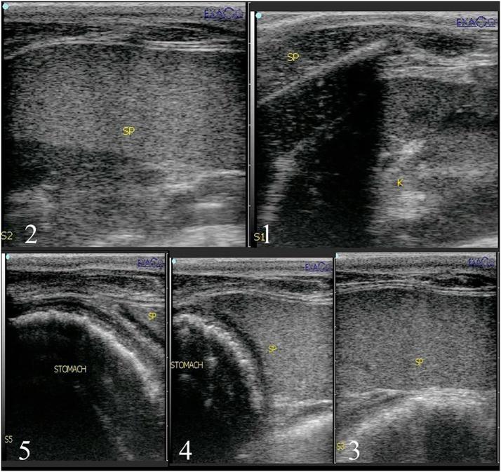
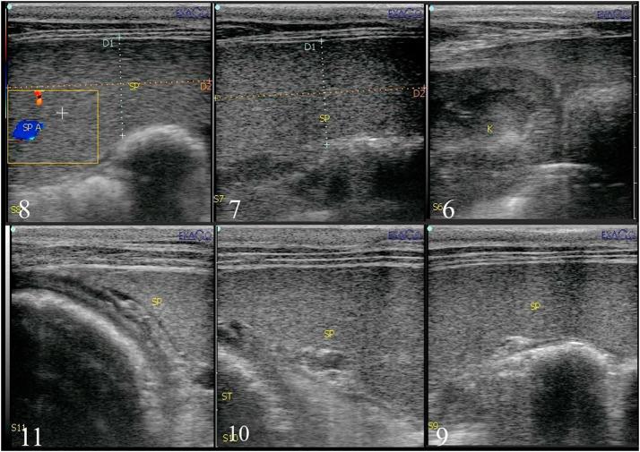
There are varieties of surgical approaches reported for equine splenectomy and all of them were dealing with the most reachable situation of splenic hilus and easy handling of the spleen. The aim of this work was to establish the normal ultrasound parameters of spleen in donkeys (normal echogenicity, hilus situation, topographic location and correlation with neighboring organs) as a guide to select the best approach for total splenectomy in donkeys. Splenic ultrasound was carried out on six normal donkeys before experimental total splenectomy in the standing position. The splenic topographic location was recorded among 4 rows including 30 squares. These animals were divided into two groups according to the surgical approach of total splenectomy. Total splenectomy after left 16 and 17 ribs partial resection in standing position was carried out in group1 and group 2, respectively. Ultrasonographically, the spleen had homogenously echogenic pattern and appeared hyperechoic to the liver. Only one third of the spleen was located in front of the 16 rib where the hilus and splenic blood vessels were nearly under the 16 rib. The splenic artery and splenic vein were ultrasonographically visualized between the left 16 and 17 ribs 10-15 cm from dorsal midline. This area was the site of the important ligation during total splenectomy. In conclusion, ultrasonography guidance for total splenectomy in donkeys assisted the surgical findings and proved that technique following partial resection of the 17 rib at the standing position is the most convenient surgical approach for total splenectomy in donkeys.
已有多种用于马脾切除术的手术方法被报道,所有这些方法都针对脾门最易触及的情况以及便于处理脾脏。本研究的目的是确定驴脾脏的正常超声参数(正常回声、脾门情况、地形位置以及与邻近器官的相关性),作为选择驴全脾切除术最佳方法的指南。在对六只正常驴进行实验性全脾切除术之前,在站立位对其进行脾脏超声检查。脾脏的地形位置记录在包括30个方格的4行中。根据全脾切除术的手术方法将这些动物分为两组。第一组和第二组分别在站立位进行左第16和17肋部分切除术后行全脾切除术。超声检查显示,脾脏具有均匀的回声模式,相对于肝脏呈高回声。只有三分之一的脾脏位于第16肋前方,脾门和脾血管几乎在第16肋下方。脾动脉和脾静脉在超声下可在距背中线10 - 15厘米的左第16和17肋之间显示。该区域是全脾切除术中重要结扎部位。总之,超声引导下的驴全脾切除术有助于手术发现,并证明在站立位切除第17肋后进行的技术是驴全脾切除术最便捷的手术方法。