Clark M W, Goelz S, Abelson J
Division of Biology, California Institute of Technology, Pasadena 91125.
EMBO J. 1988 Dec 1;7(12):3829-36. doi: 10.1002/j.1460-2075.1988.tb03268.x.
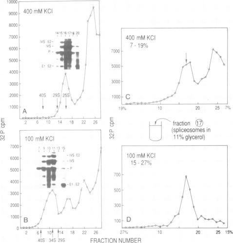
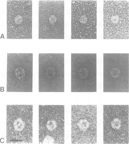
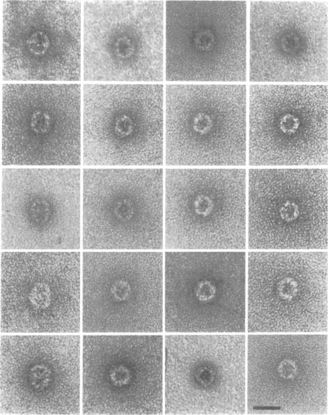
We have partially purified the yeast spliceosome by differential sedimentation in glycerol gradients. By electron microscopy we have identified a particle in these fractions that is the spliceosome. In 100 mM KCl buffer, the yeast spliceosome is an ovoid disc with the dimensions of 20 x 23.5 nm with a central indentation. To verify that these ovoid particles were spliceosomes, specific labels were used to tag them. These tagged spliceosomes were then identified in the electron microscope. The salt dependent shift of sedimentation rate for the spliceosome can be explained by a change in size of the particle.
我们通过在甘油梯度中进行差速沉降,对酵母剪接体进行了部分纯化。通过电子显微镜,我们在这些组分中鉴定出了一种颗粒,即剪接体。在100 mM KCl缓冲液中,酵母剪接体是一个椭圆形圆盘,尺寸为20×23.5 nm,中间有凹陷。为了验证这些椭圆形颗粒是剪接体,我们使用了特异性标记对它们进行标记。然后在电子显微镜下鉴定这些标记的剪接体。剪接体沉降速率的盐依赖性变化可以通过颗粒大小的改变来解释。