• 文献检索
  • 文档翻译
  • 深度研究
  • 学术资讯
  • Suppr Zotero 插件Zotero 插件
  • 邀请有礼
  • 套餐&价格
  • 历史记录
应用&插件
Suppr Zotero 插件Zotero 插件浏览器插件Mac 客户端Windows 客户端微信小程序
定价
高级版会员购买积分包购买API积分包
服务
文献检索文档翻译深度研究API 文档MCP 服务
关于我们
关于 Suppr公司介绍联系我们用户协议隐私条款
关注我们

Suppr 超能文献

核心技术专利:CN118964589B侵权必究
粤ICP备2023148730 号-1Suppr @ 2026

文献检索

告别复杂PubMed语法,用中文像聊天一样搜索,搜遍4000万医学文献。AI智能推荐,让科研检索更轻松。

立即免费搜索

文件翻译

保留排版,准确专业,支持PDF/Word/PPT等文件格式,支持 12+语言互译。

免费翻译文档

深度研究

AI帮你快速写综述,25分钟生成高质量综述,智能提取关键信息,辅助科研写作。

立即免费体验

利用锰增强 MRI 评估大鼠产前辐射诱导的海马异常。

Prenatal Irradiation-Induced Hippocampal Abnormalities in Rats Evaluated Using Manganese-Enhanced MRI.

机构信息

Division of Health Sciences, Department of Medical Physics and Engineering, Graduate School of Medicine, Osaka University, Osaka, Japan.

Department of Nutrition, Faculty of Medical and Health Sciences, Tsukuba International University, Tsuchiura, Japan.

出版信息

Front Neural Circuits. 2018 Dec 17;12:112. doi: 10.3389/fncir.2018.00112. eCollection 2018.

DOI:10.3389/fncir.2018.00112
PMID:30618648
原文链接:https://pmc.ncbi.nlm.nih.gov/articles/PMC6304475/
Abstract

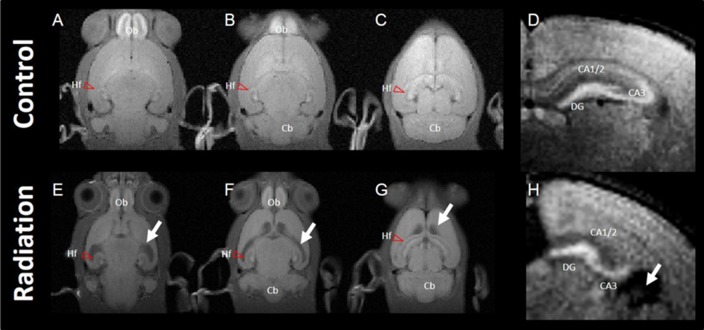
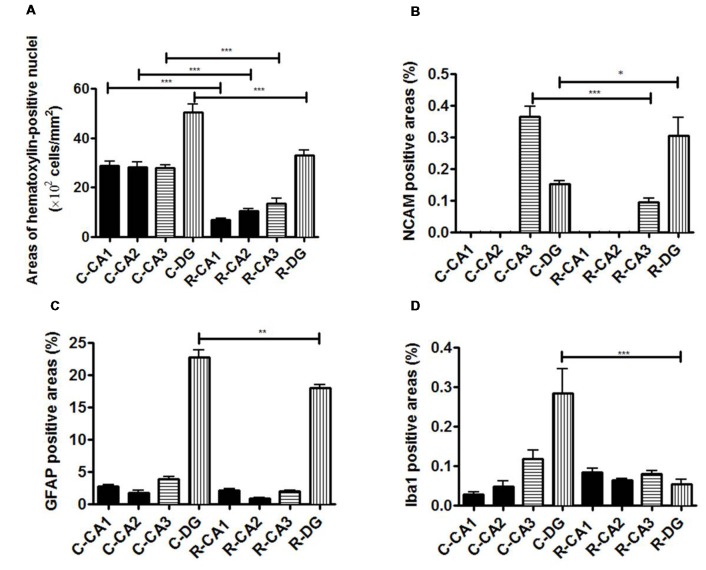
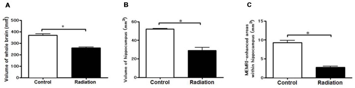
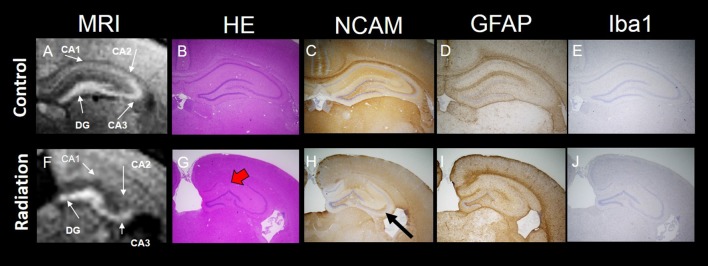
The aim of this study was to characterize hippocampal abnormalities in rats after prenatal x-ray irradiation using manganese-enhanced MRI (MEMRI). All radiation-exposed rat brains showed a reduced volume with prominent dilatation of lateral ventricles. Moreover, MEMRI-enhanced areas within the hippocampus were reduced in volumes by approximately 25% of controls, although the entire volume of hippocampus was decreased by approximately 50% of controls. MEMRI signals were enhanced strongly in the hilus and granular layer of the dentate gyrus (DG) and the pyramidal layer and infrapyramidal region of the CA3 region, and moderately along the CA1/2 pyramidal cell layer in the control rats. In radiation-exposed rats, MEMRI signals in the CA1/2 regions disappeared due to disrupting their laminar organization, although strong MEMRI signals were sustained in the DG and CA3 regions. Histopathological examinations in radiation-exposed rats revealed disorganizations of the DG granule cell layer and the CA3 pyramidal cell layer with reducing the cell density. The CA1/2 pyramidal cell layer was disrupted by invading ectopic cell mass. Neural cell adhesion molecule (NCAM)-positive fiber bundles were sustained in radiation-exposed rats, although they distributed aberrantly in the suprapyramidal CA3 region with a slight reduction of NCAM staining. Furthermore, glial components consisted largely by astrocytes and minor by microglia were densely distributed in the DG rather than in other hippocampal regions, and their density radiation-exposed rats. In conclusion, MEMRI signal enhancements could delineate different neuronal and/or glial components among hippocampal regions. We characterized microstructures of the deformed hippocampus as well as its macrostructures in a prenatally radiation-exposed rat model using MEMRI. The present findings provide advantageous information for detecting nondestructively hippocampal deformations in neurodevelopmental disorders.

摘要

本研究旨在利用锰增强磁共振成像(MEMRI)对产前 X 射线照射后的大鼠海马异常进行特征描述。所有受辐射的大鼠大脑显示出体积缩小,侧脑室明显扩张。此外,尽管海马体的整个体积减少了约 50%,但 MEMRI 增强区域的体积减少了约 25%。MEMRI 信号在齿状回(DG)的颗粒层和锥体细胞层以及 CA3 区域的锥体细胞层和下锥体细胞区强烈增强,在对照组大鼠的 CA1/2 锥体细胞层适度增强。在受辐射的大鼠中,由于层状结构破坏,CA1/2 区的 MEMRI 信号消失,尽管 DG 和 CA3 区仍保持强烈的 MEMRI 信号。受辐射大鼠的组织病理学检查显示 DG 颗粒细胞层和 CA3 锥体细胞层排列紊乱,细胞密度降低。CA1/2 锥体细胞层被异位细胞团破坏。神经细胞黏附分子(NCAM)阳性纤维束在受辐射的大鼠中得以维持,尽管它们在 CA3 上区的分布异常,NCAM 染色略有减少。此外,胶质细胞成分主要由星形胶质细胞和少量小胶质细胞组成,在 DG 中分布密集,而不是在其他海马区,其密度在受辐射的大鼠中增加。总之,MEMRI 信号增强可以描绘出海马区域中不同的神经元和/或神经胶质成分。我们使用 MEMRI 对产前辐射暴露大鼠模型中的变形海马体的微观结构及其宏观结构进行了特征描述。这些发现为非破坏性检测神经发育障碍中海马体变形提供了有益信息。

https://cdn.ncbi.nlm.nih.gov/pmc/blobs/de7d/6304475/bfe7e7c7c97c/fncir-12-00112-g0004.jpg
https://cdn.ncbi.nlm.nih.gov/pmc/blobs/de7d/6304475/872b39df2e99/fncir-12-00112-g0001.jpg
https://cdn.ncbi.nlm.nih.gov/pmc/blobs/de7d/6304475/7a0fb5cae947/fncir-12-00112-g0002.jpg
https://cdn.ncbi.nlm.nih.gov/pmc/blobs/de7d/6304475/eef6bc122d7c/fncir-12-00112-g0003.jpg
https://cdn.ncbi.nlm.nih.gov/pmc/blobs/de7d/6304475/bfe7e7c7c97c/fncir-12-00112-g0004.jpg

相似文献

1
Prenatal Irradiation-Induced Hippocampal Abnormalities in Rats Evaluated Using Manganese-Enhanced MRI.利用锰增强 MRI 评估大鼠产前辐射诱导的海马异常。
Front Neural Circuits. 2018 Dec 17;12:112. doi: 10.3389/fncir.2018.00112. eCollection 2018.
2
Manganese-enhanced MRI reflects seizure outcome in a model for mesial temporal lobe epilepsy.锰增强 MRI 反映内侧颞叶癫痫模型中的癫痫发作结局。
Neuroimage. 2013 Mar;68:30-8. doi: 10.1016/j.neuroimage.2012.11.054. Epub 2012 Dec 5.
3
Quantitative assessment of central nervous system disorder induced by prenatal X-ray exposure using diffusion and manganese-enhanced MRI.采用弥散加权和锰增强 MRI 对产前 X 射线照射致中枢神经系统障碍进行定量评估。
NMR Biomed. 2012 Jan;25(1):75-83. doi: 10.1002/nbm.1715. Epub 2011 Apr 28.
4
Manganese enhanced MRI detects mossy fiber sprouting rather than neurodegeneration, gliosis or seizure-activity in the epileptic rat hippocampus.锰增强磁共振成像检测到癫痫大鼠海马体中的苔藓纤维发芽,而非神经退行性变、胶质增生或癫痫活动。
Neuroimage. 2008 May 1;40(4):1718-30. doi: 10.1016/j.neuroimage.2008.01.042. Epub 2008 Feb 7.
5
Hippocampal changes in inflammasomes, apoptosis, and MEMRI after radiation-induced brain injury in juvenile rats.幼年大鼠辐射性脑损伤后海马中炎性小体、细胞凋亡及磁共振弹性成像的变化
Radiat Oncol. 2020 Apr 10;15(1):78. doi: 10.1186/s13014-020-01525-3.
6
Effect of prenatal protein malnutrition on numbers of neurons in the principal cell layers of the adult rat hippocampal formation.产前蛋白质营养不良对成年大鼠海马结构主要细胞层中神经元数量的影响。
Hippocampus. 2005;15(3):393-403. doi: 10.1002/hipo.20065.
7
Multiparametric MRI evaluation of kainic acid-induced neuronal activation in rat hippocampus.对大鼠海马体中红藻氨酸诱导的神经元激活的多参数磁共振成像评估
Brain. 2007 Dec;130(Pt 12):3124-34. doi: 10.1093/brain/awm207. Epub 2007 Sep 4.
8
Cellular and connective organization of slice cultures of the rat hippocampus and fascia dentata.大鼠海马体和齿状回切片培养物的细胞与结缔组织结构
J Comp Neurol. 1984 Sep 20;228(3):432-46. doi: 10.1002/cne.902280310.
9
Differential Local Connectivity and Neuroinflammation Profiles in the Medial Prefrontal Cortex and Hippocampus in the Valproic Acid Rat Model of Autism.丙戊酸诱导的自闭症大鼠模型中内侧前额叶皮质和海马的差异局部连接性及神经炎症特征
Dev Neurosci. 2015;37(3):215-31. doi: 10.1159/000375489. Epub 2015 Apr 18.
10
Prenatal gonadal steroids affect adult spatial behavior, CA1 and CA3 pyramidal cell morphology in rats.产前性腺类固醇会影响成年大鼠的空间行为、海马CA1区和CA3区锥体细胞形态。
Horm Behav. 1998 Oct;34(2):183-98. doi: 10.1006/hbeh.1998.1477.

引用本文的文献

1
Comparative Analysis of Behavioral Reactions and Morphological Changes in the Rat Brain After Exposure to Ionizing Radiation with Different Physical Characteristics.不同物理特性电离辐射暴露后大鼠大脑行为反应与形态学变化的比较分析
Cell Mol Neurobiol. 2023 Jan;43(1):339-353. doi: 10.1007/s10571-021-01187-z. Epub 2022 Jan 4.
2
Hippocampal changes in inflammasomes, apoptosis, and MEMRI after radiation-induced brain injury in juvenile rats.幼年大鼠辐射性脑损伤后海马中炎性小体、细胞凋亡及磁共振弹性成像的变化
Radiat Oncol. 2020 Apr 10;15(1):78. doi: 10.1186/s13014-020-01525-3.
3
Manganese-Enhanced Magnetic Resonance Imaging: Application in Central Nervous System Diseases.

本文引用的文献

1
Early detection of elevated lactate levels in a mitochondrial disease model using chemical exchange saturation transfer (CEST) and magnetic resonance spectroscopy (MRS) at 7T-MRI.在7T磁共振成像(MRI)中,利用化学交换饱和转移(CEST)和磁共振波谱(MRS)在一种线粒体疾病模型中早期检测乳酸水平升高。
Radiol Phys Technol. 2019 Mar;12(1):46-54. doi: 10.1007/s12194-018-0490-1. Epub 2018 Nov 22.
2
Dose-related cerebellar abnormality in rats with prenatal exposure to X-irradiation by magnetic resonance imaging volumetric analysis.通过磁共振成像体积分析对产前暴露于X射线的大鼠进行剂量相关的小脑异常研究。
Congenit Anom (Kyoto). 2013 Sep;53(3):127-30. doi: 10.1111/cga.12016.
3
锰增强磁共振成像:在中枢神经系统疾病中的应用
Front Neurol. 2020 Feb 25;11:143. doi: 10.3389/fneur.2020.00143. eCollection 2020.
Manganese enhanced MRI detects mossy fiber sprouting rather than neurodegeneration, gliosis or seizure-activity in the epileptic rat hippocampus.
锰增强磁共振成像检测到癫痫大鼠海马体中的苔藓纤维发芽,而非神经退行性变、胶质增生或癫痫活动。
Neuroimage. 2008 May 1;40(4):1718-30. doi: 10.1016/j.neuroimage.2008.01.042. Epub 2008 Feb 7.
4
Manganese-enhanced magnetic resonance imaging of mossy fiber plasticity in vivo.体内苔藓纤维可塑性的锰增强磁共振成像
Neuroimage. 2006 Mar;30(1):130-5. doi: 10.1016/j.neuroimage.2005.09.007. Epub 2005 Oct 24.
5
Effects of prenatal irradiation with an accelerated heavy-ion beam on postnatal development in rats: I. Neurophysiological alterations.加速重离子束产前照射对大鼠出生后发育的影响:I. 神经生理学改变
Radiat Res. 2005 Oct;164(4 Pt 2):561-6. doi: 10.1667/rr3334.1.
6
Ectopic neurons in the hippocampus may be a cause of learning disability after prenatal exposure to X-rays in rats.
J Radiat Res. 2004 Dec;45(4):563-9. doi: 10.1269/jrr.45.563.
7
Functional mapping of neural pathways in rodent brain in vivo using manganese-enhanced three-dimensional magnetic resonance imaging.利用锰增强三维磁共振成像对啮齿动物大脑中的神经通路进行体内功能映射。
NMR Biomed. 2004 Dec;17(8):554-68. doi: 10.1002/nbm.937.
8
Manganese-enhanced magnetic resonance imaging (MEMRI): methodological and practical considerations.锰增强磁共振成像(MEMRI):方法学与实际考量
NMR Biomed. 2004 Dec;17(8):532-43. doi: 10.1002/nbm.945.
9
In vivo detection of neuroarchitecture in the rodent brain using manganese-enhanced MRI.利用锰增强磁共振成像在啮齿动物大脑中进行神经结构的体内检测。
Neuroimage. 2004 Jul;22(3):1046-59. doi: 10.1016/j.neuroimage.2004.03.031.
10
The effect of the timing of prenatal X-irradiation on Purkinje cell numbers in rat cerebellum.产前X射线照射时间对大鼠小脑浦肯野细胞数量的影响。
Brain Res Dev Brain Res. 2002 Dec 15;139(2):159-66. doi: 10.1016/s0165-3806(02)00542-4.