Herminghaus Anna, Papenbrock Henrike, Eberhardt Rebecca, Vollmer Christian, Truse Richard, Schulz Jan, Bauer Inge, Weidinger Adelheid, Kozlov Andrey V, Stiban Johnny, Picker Olaf
Department of Anaesthesiology, University of Düsseldorf, Moorenstrasse 5, 40225, Düsseldorf, Germany.
Ludwig Boltzmann Institute for Clinical and Experimental Traumatology, AUVA Research Center, Donaueschingenstraße 13, 1200, Wien, Austria.
Intensive Care Med Exp. 2019 Jan 8;7(1):4. doi: 10.1186/s40635-018-0219-9.
Evidence suggests that early adaptive responses of hepatic mitochondria occur in experimentally induced sepsis. Little is known about both colonic mitochondrial function during abdominal infection and long-term changes in mitochondrial function under inflammatory conditions. We hypothesize that hepatic and colonic mitochondrial oxygen consumption changes time-dependently after sterile laparotomy and in the course of abdominal infection. The aim of the present study was to investigate the hepatic and colonic mitochondrial respiration after sterile laparotomy and abdominal infection over up to 96 h.
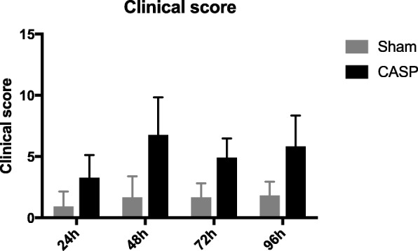
After approval of the local Animal Care and Use Committee, 95 Wistar rats were randomized into 8 groups (n = 11-12): 1-4 sham (laparotomy only) and 5-8 colon ascendens stent peritonitis (CASP). Healthy, unoperated animals served as controls (n = 9). The mitochondrial respiration in colon and liver homogenates was assessed 24, 48, 72, and 96 h after surgery. Mitochondrial oxygen consumption was determined using a Clark-type electrode. State 2 (oxygen consumption in the presence of the substrates for complexes I and II) and state 3 respiration (ADP dependent) were assessed. The respiratory control ratio (RCR state 3/state 2) and ADP/O ratio (ADP added/oxygen consumed) were calculated for both complexes. Data are presented as means ± SD, two-way ANOVA followed by Tukey's post hoc test.
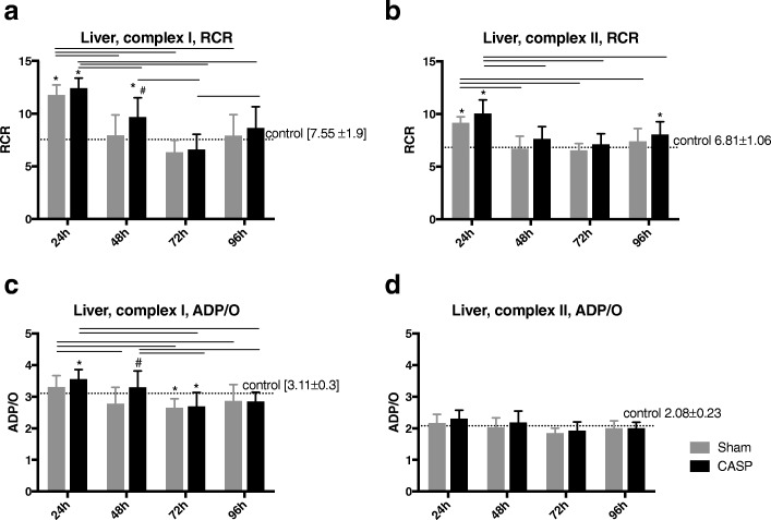
Hepatic RCR was initially (after 24 h) elevated in both operated groups; after 48 h only, the septic group was elevated compared to controls. In CASP groups, the hepatic ADP/O ratio for complex I was elevated after 24 h (vs. controls) and after 48 h (vs. sham) but declined after 72 h (vs. controls). The ADP/O ratio for complex II stayed unchanged over the time period until 96 h. The colonic RCR and ADP/O did not change over time after sham or CASP operation.
Hepatic, but not colonic, mitochondrial respiration is increased in the initial phase (until 48 h) and normalizes in the longer course of time (until 96 h) of abdominal infection.
有证据表明,在实验性诱导的脓毒症中,肝线粒体的早期适应性反应会出现。关于腹部感染期间结肠线粒体功能以及炎症条件下线粒体功能的长期变化,人们了解甚少。我们推测,在无菌剖腹术后以及腹部感染过程中,肝和结肠线粒体的氧消耗会随时间而变化。本研究的目的是调查无菌剖腹术和腹部感染后长达96小时内肝和结肠的线粒体呼吸情况。
经当地动物护理和使用委员会批准后,将95只Wistar大鼠随机分为8组(每组n = 11 - 12只):1 - 4组为假手术组(仅剖腹术),5 - 8组为升结肠支架性腹膜炎(CASP)组。健康未手术的动物作为对照组(n = 9只)。在术后24、48、72和96小时评估结肠和肝匀浆中的线粒体呼吸。使用Clark型电极测定线粒体氧消耗。评估状态2(存在复合物I和II的底物时的氧消耗)和状态3呼吸(依赖ADP)。计算两种复合物的呼吸控制率(RCR状态3/状态2)和ADP/O比率(添加的ADP/消耗的氧)。数据以均值±标准差表示,采用双向方差分析,随后进行Tukey事后检验。
两个手术组的肝RCR最初(24小时后)均升高;仅在48小时后,脓毒症组相对于对照组升高。在CASP组中,复合物I的肝ADP/O比率在24小时后(与对照组相比)和48小时后(与假手术组相比)升高,但在72小时后(与对照组相比)下降。复合物II的ADP/O比率在直至96小时的时间段内保持不变。假手术或CASP手术后,结肠的RCR和ADP/O随时间未发生变化。
在腹部感染的初始阶段(直至48小时),肝线粒体呼吸增加,而结肠线粒体呼吸未增加,在较长时间(直至96小时)内肝线粒体呼吸恢复正常。