• 文献检索
  • 文档翻译
  • 深度研究
  • 学术资讯
  • Suppr Zotero 插件Zotero 插件
  • 邀请有礼
  • 套餐&价格
  • 历史记录
应用&插件
Suppr Zotero 插件Zotero 插件浏览器插件Mac 客户端Windows 客户端微信小程序
定价
高级版会员购买积分包购买API积分包
服务
文献检索文档翻译深度研究API 文档MCP 服务
关于我们
关于 Suppr公司介绍联系我们用户协议隐私条款
关注我们

Suppr 超能文献

核心技术专利:CN118964589B侵权必究
粤ICP备2023148730 号-1Suppr @ 2026

文献检索

告别复杂PubMed语法,用中文像聊天一样搜索,搜遍4000万医学文献。AI智能推荐,让科研检索更轻松。

立即免费搜索

文件翻译

保留排版,准确专业,支持PDF/Word/PPT等文件格式,支持 12+语言互译。

免费翻译文档

深度研究

AI帮你快速写综述,25分钟生成高质量综述,智能提取关键信息,辅助科研写作。

立即免费体验

MR 细胞外容积成像和非对比 T1ρ 成像可早期检测糖尿病猴心肌纤维化。

MR extracellular volume mapping and non-contrast T1ρ mapping allow early detection of myocardial fibrosis in diabetic monkeys.

机构信息

Department of Radiology, West China Hospital, Sichuan University, 37 Guoxue Alley, Chengdu, 610041, Sichuan, China.

Sichuan Primed Shines Bio-tech Co., Ltd., Chengdu, China.

出版信息

Eur Radiol. 2019 Jun;29(6):3006-3016. doi: 10.1007/s00330-018-5950-9. Epub 2019 Jan 14.

DOI:10.1007/s00330-018-5950-9
PMID:30643944
原文链接:https://pmc.ncbi.nlm.nih.gov/articles/PMC6510861/
Abstract

OBJECTIVE

To detect diffuse myocardial fibrosis in different severity levels of left ventricular diastolic dysfunction (DD) in spontaneous type 2 diabetes mellitus (T2DM) rhesus monkeys.

METHODS

Eighteen spontaneous T2DM and nine healthy monkeys were studied. Echocardiography was performed for diastolic function classification. Cardiac magnetic resonance (CMR) imaging was performed to obtain extracellular volume fraction (ECV) maps and T1ρ maps at two different spin-locking frequencies. ECV values, T1ρ values, and myocardial fibrosis index (mFI) values which are based on the dispersion of T1ρ, were calculated. Global peak diastolic longitudinal strain rates (GSrL) were also obtained.

RESULTS

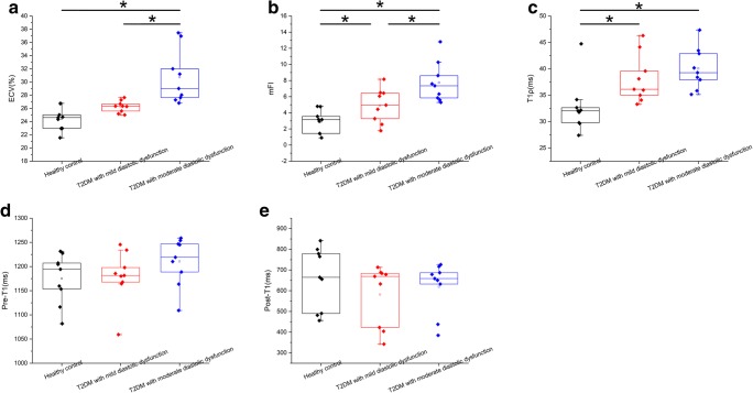
Echocardiography results showed mild DD in nine T2DM monkeys and moderate DD in the other nine. The global ECV values were significantly different among healthy animals as compared with animals with mild DD or moderate DD, and the ECV values of animals with moderate DD were significantly higher as compared with those of mild DD. The mFI values increased progressively from healthy animals to those with mild DD and then to those with moderate DD. Diastolic function indicators (e.g., early diastolic mitral annulus velocity, GSrL) correlated well with ECV and mFI.

CONCLUSIONS

Monkeys with T2DM exhibit increased ECV, T1ρ, and mFI values, which may be indicative of the expansion of extracellular volume and the deposition of excessive collagen. T1ρ mapping may have the potential to be used for diffuse myocardial fibrosis assessment.

KEY POINTS

• Monkeys with T2DM exhibit increased ECV, T1ρ, and mFI values, which may be indicative of the expansion of extracellular volume and the deposition of excessive collagen. • The relationship between diastolic dysfunction and diffuse myocardial fibrosis may be demonstrated by imaging markers. • Non-contrast T1ρ mapping may have the potential to be used for diffuse myocardial assessment.

摘要

目的

检测不同严重程度左心室舒张功能障碍(DD)的自发性 2 型糖尿病(T2DM)恒河猴的弥漫性心肌纤维化。

方法

研究了 18 只自发性 T2DM 猴和 9 只健康猴。行超声心动图检查进行舒张功能分类。行心脏磁共振(CMR)成像以获得细胞外容积分数(ECV)图和两个不同自旋锁定频率的 T1ρ 图。计算 ECV 值、T1ρ 值和基于 T1ρ 离散的心肌纤维化指数(mFI)值。还获得了整体峰值舒张纵向应变率(GSrL)。

结果

超声心动图结果显示,9 只 T2DM 猴存在轻度 DD,另外 9 只存在中度 DD。与轻度 DD 或中度 DD 的动物相比,健康动物的整体 ECV 值有显著差异,中度 DD 动物的 ECV 值显著高于轻度 DD 动物。mFI 值从健康动物逐渐升高至轻度 DD 动物,然后再升高至中度 DD 动物。舒张功能指标(如早期舒张二尖瓣环速度、GSrL)与 ECV 和 mFI 相关性良好。

结论

T2DM 猴存在 ECV、T1ρ 和 mFI 值升高,可能提示细胞外容积扩张和胶原过度沉积。T1ρ 图可能具有评估弥漫性心肌纤维化的潜力。

关键点

• T2DM 猴存在 ECV、T1ρ 和 mFI 值升高,可能提示细胞外容积扩张和胶原过度沉积。

• 舒张功能障碍与弥漫性心肌纤维化之间的关系可以通过影像学标志物来证明。

• 无需对比剂的 T1ρ 图可能具有评估弥漫性心肌的潜力。

https://cdn.ncbi.nlm.nih.gov/pmc/blobs/ecd7/6510861/b1e235edbe50/330_2018_5950_Fig5_HTML.jpg
https://cdn.ncbi.nlm.nih.gov/pmc/blobs/ecd7/6510861/c0f6ea7d072e/330_2018_5950_Fig1_HTML.jpg
https://cdn.ncbi.nlm.nih.gov/pmc/blobs/ecd7/6510861/83a4da4ebd6c/330_2018_5950_Fig2_HTML.jpg
https://cdn.ncbi.nlm.nih.gov/pmc/blobs/ecd7/6510861/0413263f4e8d/330_2018_5950_Fig3_HTML.jpg
https://cdn.ncbi.nlm.nih.gov/pmc/blobs/ecd7/6510861/ad62bbb2af66/330_2018_5950_Fig4_HTML.jpg
https://cdn.ncbi.nlm.nih.gov/pmc/blobs/ecd7/6510861/b1e235edbe50/330_2018_5950_Fig5_HTML.jpg

相似文献

1
MR extracellular volume mapping and non-contrast T1ρ mapping allow early detection of myocardial fibrosis in diabetic monkeys.MR 细胞外容积成像和非对比 T1ρ 成像可早期检测糖尿病猴心肌纤维化。
Eur Radiol. 2019 Jun;29(6):3006-3016. doi: 10.1007/s00330-018-5950-9. Epub 2019 Jan 14.
2
Noncontrast T1ρ dispersion imaging is sensitive to diffuse fibrosis: A cardiovascular magnetic resonance study at 3T in hypertrophic cardiomyopathy.非对比 T1ρ 弥散成像对弥漫性纤维化敏感:肥厚型心肌病的 3T 心血管磁共振研究。
Magn Reson Imaging. 2022 Sep;91:1-8. doi: 10.1016/j.mri.2022.05.001. Epub 2022 May 4.
3
Early detection of myocardial involvement by non-contrast T1ρ mapping of cardiac magnetic resonance in type 2 diabetes mellitus.2 型糖尿病患者心脏磁共振钆对比 T1ρ mapping 早期检测心肌受累。
Front Endocrinol (Lausanne). 2024 Mar 5;15:1335899. doi: 10.3389/fendo.2024.1335899. eCollection 2024.
4
2D/3D CMR tissue tracking versus CMR tagging in the assessment of spontaneous T2DM rhesus monkeys with isolated diastolic dysfunction.在评估自发性2型糖尿病且伴有单纯舒张功能障碍的恒河猴时,二维/三维心脏磁共振成像组织追踪与心脏磁共振标记技术的比较
BMC Med Imaging. 2018 Nov 26;18(1):47. doi: 10.1186/s12880-018-0288-y.
5
Cardiac magnetic resonance T1 mapping for evaluating myocardial fibrosis in patients with type 2 diabetes mellitus: correlation with left ventricular longitudinal diastolic dysfunction.心脏磁共振 T1 mapping 评估 2 型糖尿病患者心肌纤维化:与左心室长轴舒张功能障碍的相关性。
Eur Radiol. 2022 Nov;32(11):7647-7656. doi: 10.1007/s00330-022-08800-9. Epub 2022 May 14.
6
Myocardial fibrosis imaging based on T1-mapping and extracellular volume fraction (ECV) measurement in muscular dystrophy patients: diagnostic value compared with conventional late gadolinium enhancement (LGE) imaging.基于T1映射和细胞外容积分数(ECV)测量的肌营养不良患者心肌纤维化成像:与传统钆延迟增强(LGE)成像相比的诊断价值
Eur Heart J Cardiovasc Imaging. 2014 Sep;15(9):1004-12. doi: 10.1093/ehjci/jeu050. Epub 2014 Mar 30.
7
Extracellular volume fraction is more closely associated with altered regional left ventricular velocities than left ventricular ejection fraction in nonischemic cardiomyopathy.在非缺血性心肌病中,细胞外容积分数比左心室射血分数与左心室局部速度改变的相关性更强。
Circ Cardiovasc Imaging. 2014 Dec 31;8(1). doi: 10.1161/CIRCIMAGING.114.001998. Print 2015 Jan.
8
Diastolic dysfunction in spontaneous type 2 diabetes rhesus monkeys: a study using echocardiography and magnetic resonance imaging.自发性2型糖尿病恒河猴的舒张功能障碍:一项使用超声心动图和磁共振成像的研究。
BMC Cardiovasc Disord. 2015 Jun 26;15:59. doi: 10.1186/s12872-015-0046-9.
9
Dynamic changes in cardiac morphology, function, and diffuse myocardial fibrosis duration of diabetes in type 1 and type 2 diabetic mice models using 7.0 T CMR and echocardiography.使用 7.0T CMR 和超声心动图观察 1 型和 2 型糖尿病小鼠模型中心脏形态、功能和弥漫性心肌纤维化持续时间的动态变化。
Front Endocrinol (Lausanne). 2023 Nov 8;14:1278619. doi: 10.3389/fendo.2023.1278619. eCollection 2023.
10
Free-breathing non-contrast T1ρ dispersion magnetic resonance imaging of myocardial interstitial fibrosis in comparison with extracellular volume fraction.自由呼吸非对比T1ρ弥散磁共振成像评估心肌间质纤维化与细胞外容积分数的比较
J Cardiovasc Magn Reson. 2024;26(2):101093. doi: 10.1016/j.jocmr.2024.101093. Epub 2024 Sep 6.

引用本文的文献

1
Endogenous assessment of late gadolinium enhancement grey area in patients with chronic myocardial infarction using T1rho mapping.使用T1rho成像对慢性心肌梗死患者晚期钆增强灰色区域进行内源性评估。
Quant Imaging Med Surg. 2025 Aug 1;15(8):7183-7194. doi: 10.21037/qims-24-1703. Epub 2025 Jul 30.
2
Multiparametric quantitative magnetic resonance imaging of skeletal muscle in CKD.慢性肾脏病患者骨骼肌的多参数定量磁共振成像
Am J Physiol Renal Physiol. 2025 Jul 1;329(1):F99-F111. doi: 10.1152/ajprenal.00254.2024. Epub 2025 May 27.
3
Supaglutide alleviates hepatic steatosis in monkeys with spontaneous MASH.

本文引用的文献

1
Heart Failure With Preserved Ejection Fraction in Diabetes: Mechanisms and Management.糖尿病合并射血分数保留的心力衰竭:机制与治疗。
Can J Cardiol. 2018 May;34(5):632-643. doi: 10.1016/j.cjca.2018.02.026. Epub 2018 Mar 2.
2
Multiple common comorbidities produce left ventricular diastolic dysfunction associated with coronary microvascular dysfunction, oxidative stress, and myocardial stiffening.多种常见合并症导致左心室舒张功能障碍,与冠状动脉微血管功能障碍、氧化应激和心肌僵硬有关。
Cardiovasc Res. 2018 Jun 1;114(7):954-964. doi: 10.1093/cvr/cvy038.
3
Increased myocardial extracellular volume assessed by cardiovascular magnetic resonance T1 mapping and its determinants in type 2 diabetes mellitus patients with normal myocardial systolic strain.
司美格鲁肽可减轻自发性代谢相关脂肪性肝病(MASH)猴子的肝脂肪变性。
Diabetol Metab Syndr. 2024 Dec 19;16(1):303. doi: 10.1186/s13098-024-01513-7.
4
Free-breathing non-contrast T1ρ dispersion magnetic resonance imaging of myocardial interstitial fibrosis in comparison with extracellular volume fraction.自由呼吸非对比T1ρ弥散磁共振成像评估心肌间质纤维化与细胞外容积分数的比较
J Cardiovasc Magn Reson. 2024;26(2):101093. doi: 10.1016/j.jocmr.2024.101093. Epub 2024 Sep 6.
5
Role of endogenous T1ρ and its dispersion imaging in differential diagnosis of cardiac amyloidosis.内源性T1ρ及其弥散成像在心脏淀粉样变性鉴别诊断中的作用。
J Cardiovasc Magn Reson. 2024;26(2):101080. doi: 10.1016/j.jocmr.2024.101080. Epub 2024 Aug 8.
6
Early detection of myocardial involvement by non-contrast T1ρ mapping of cardiac magnetic resonance in type 2 diabetes mellitus.2 型糖尿病患者心脏磁共振钆对比 T1ρ mapping 早期检测心肌受累。
Front Endocrinol (Lausanne). 2024 Mar 5;15:1335899. doi: 10.3389/fendo.2024.1335899. eCollection 2024.
7
Dynamic changes in cardiac morphology, function, and diffuse myocardial fibrosis duration of diabetes in type 1 and type 2 diabetic mice models using 7.0 T CMR and echocardiography.使用 7.0T CMR 和超声心动图观察 1 型和 2 型糖尿病小鼠模型中心脏形态、功能和弥漫性心肌纤维化持续时间的动态变化。
Front Endocrinol (Lausanne). 2023 Nov 8;14:1278619. doi: 10.3389/fendo.2023.1278619. eCollection 2023.
8
Cardiovascular magnetic resonance imaging for sequential assessment of cardiac fibrosis in mice: technical advancements and reverse translation.心血管磁共振成像用于小鼠心脏纤维化的序贯评估:技术进展和反向翻译。
Am J Physiol Heart Circ Physiol. 2024 Jan 1;326(1):H1-H24. doi: 10.1152/ajpheart.00437.2023. Epub 2023 Nov 3.
9
Imaging characterization of myocardial function, fibrosis, and perfusion in a nonhuman primate model with heart failure-like features.具有心力衰竭样特征的非人灵长类动物模型中心肌功能、纤维化和灌注的影像学特征
Front Cardiovasc Med. 2023 Aug 17;10:1214249. doi: 10.3389/fcvm.2023.1214249. eCollection 2023.
10
Robust cardiac mapping at 3T using adiabatic spin-lock preparations.在 3T 场强下使用绝热锁相准备技术实现稳健的心脏图谱绘制。
Magn Reson Med. 2023 Oct;90(4):1363-1379. doi: 10.1002/mrm.29713. Epub 2023 May 28.
心血管磁共振 T1 映射评估的心肌细胞外容积增加及其在心肌收缩应变正常的 2 型糖尿病患者中的决定因素。
Cardiovasc Diabetol. 2018 Jan 4;17(1):7. doi: 10.1186/s12933-017-0651-2.
4
Pathogenesis, Clinical Features and Treatment of Diabetic Cardiomyopathy.糖尿病心肌病的发病机制、临床特征及治疗。
Adv Exp Med Biol. 2018;1067:197-217. doi: 10.1007/5584_2017_105.
5
Native T1 Mapping and Extracellular Volume Mapping for the Assessment of Diffuse Myocardial Fibrosis in Dilated Cardiomyopathy.用于评估扩张型心肌病弥漫性心肌纤维化的 native T1 mapping 和细胞外容积 mapping。
JACC Cardiovasc Imaging. 2018 Jan;11(1):48-59. doi: 10.1016/j.jcmg.2017.04.006. Epub 2017 Jun 14.
6
A non-contrast CMR index for assessing myocardial fibrosis.一种用于评估心肌纤维化的非对比心脏磁共振成像指标。
Magn Reson Imaging. 2017 Oct;42:69-73. doi: 10.1016/j.mri.2017.04.012. Epub 2017 Apr 28.
7
The quantification of blood-brain barrier disruption using dynamic contrast-enhanced magnetic resonance imaging in aging rhesus monkeys with spontaneous type 2 diabetes mellitus.使用动态对比增强磁共振成像定量评估自发性 2 型糖尿病衰老恒河猴的血脑屏障破坏。
Neuroimage. 2017 Sep;158:480-487. doi: 10.1016/j.neuroimage.2016.07.017. Epub 2016 Jul 9.
8
Endogenous assessment of diffuse myocardial fibrosis in patients with T -mapping.T 映射技术对患者弥漫性心肌纤维化的内源性评估
J Magn Reson Imaging. 2017 Jan;45(1):132-138. doi: 10.1002/jmri.25340. Epub 2016 Jun 16.
9
Histological validation of cardiac magnetic resonance T mapping for detecting diffuse myocardial fibrosis in diabetic rabbits.心脏磁共振T成像用于检测糖尿病兔弥漫性心肌纤维化的组织学验证
J Magn Reson Imaging. 2016 Nov;44(5):1179-1185. doi: 10.1002/jmri.25268. Epub 2016 Apr 9.
10
Hyperglycemia-Induced Hypovolemia Is Involved in Early Cardiac Magnetic Resonance Alterations in Streptozotocin-Induced Diabetic Mice: A Comparison with Furosemide-Induced Hypovolemia.高血糖诱导的血容量减少参与链脲佐菌素诱导的糖尿病小鼠早期心脏磁共振改变:与呋塞米诱导的血容量减少的比较
PLoS One. 2016 Feb 22;11(2):e0149808. doi: 10.1371/journal.pone.0149808. eCollection 2016.