From the Aging Research Center (C.S.D., A.M., D.R., R.W., J.S., W.X.), Department of Neurobiology, Care Sciences and Society, Karolinska Institutet and Stockholm University; Department of Clinical Neuroscience (J.S.), Psychology Division, Karolinska Institutet, Stockholm, Sweden; Department of Biomedical Engineering (K.A.), Illinois Institute of Technology, Chicago; Rush Alzheimer's Disease Center (K.A., D.A.B.), and Department of Neurological Sciences (D.A.B.), Rush University Medical Center, Chicago, IL; and Department of Epidemiology and Biostatistics (W.X.), School of Public Health, Tianjin Medical University, China.
Neurology. 2019 Feb 12;92(7):e700-e709. doi: 10.1212/WNL.0000000000006919. Epub 2019 Jan 16.
We aimed to examine whether impaired olfaction is associated with cognitive decline and indicators of neurodegeneration in the brain of dementia-free older adults.
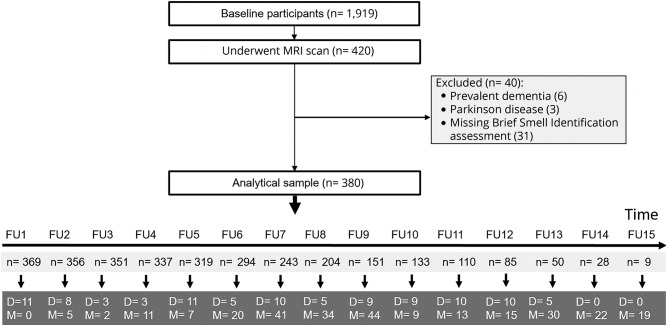
Within the Rush Memory and Aging Project, 380 dementia-free participants (mean age = 78 years) were followed for up to 15 years, and underwent MRI scans. Olfactory function was assessed using the Brief Smell Identification Test (B-SIT) at baseline, and categorized as anosmia (B-SIT <6), hyposmia (B-SIT 6-10 in men and 6-10.25 in women), and normal (B-SIT 10.25-12 in men and 10.5-12 in women). Cognitive function was annually assessed with a battery of 21 tests, from which composite scores were derived. Structural total and regional brain volumes were estimated. Data were analyzed using linear regression and mixed-effects models.
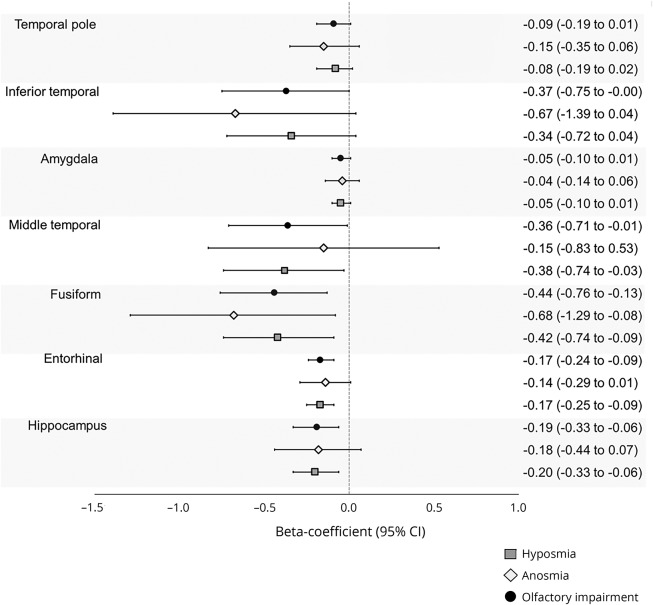
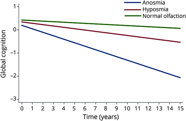
At study entry, 138 (36.3%) had normal olfactory function, 213 (56.1%) had hyposmia, and 29 (7.6%) had anosmia. In multiadjusted mixed-effects models, hyposmia (β = -0.03, 95% confidence interval [CI] -0.05 to -0.02) and anosmia (β = -0.13, 95% CI -0.16 to -0.09) were associated with faster rate of cognitive decline compared to normal olfaction. On MRI, impaired olfaction (hyposmia or anosmia) was related to smaller volumes of the hippocampus (β = -0.19, 95% CI -0.33 to -0.05), and in the entorhinal (β = -0.16, 95% CI -0.24 to -0.08), fusiform (β = -0.45, 95% CI -0.78 to -0.14), and middle temporal (β = -0.38, 95% CI -0.72 to -0.01) cortices.
Impaired olfaction predicts faster cognitive decline and might indicate neurodegeneration in the brain among dementia-free older adults.
我们旨在研究嗅觉障碍是否与痴呆症老年人的认知能力下降和大脑神经退行性变的标志物有关。
在 Rush 记忆与衰老项目中,对 380 名无痴呆症的参与者(平均年龄为 78 岁)进行了长达 15 年的随访,并进行了 MRI 扫描。在基线时使用 Brief Smell Identification Test(B-SIT)评估嗅觉功能,并分为嗅觉丧失(B-SIT <6)、嗅觉减退(男性 B-SIT 6-10,女性 B-SIT 6-10.25)和正常嗅觉(男性 B-SIT 10.25-12,女性 B-SIT 10.5-12)。认知功能每年通过一系列 21 项测试进行评估,并从中得出综合评分。估计了大脑的总结构和区域体积。使用线性回归和混合效应模型进行数据分析。
在研究开始时,138 人(36.3%)嗅觉功能正常,213 人(56.1%)嗅觉减退,29 人(7.6%)嗅觉丧失。在多因素混合效应模型中,嗅觉减退(β=-0.03,95%置信区间 [CI] -0.05 至 -0.02)和嗅觉丧失(β=-0.13,95%CI -0.16 至 -0.09)与正常嗅觉相比,认知衰退速度更快。在 MRI 上,嗅觉障碍(嗅觉减退或嗅觉丧失)与海马体体积较小有关(β=-0.19,95%CI -0.33 至 -0.05),与内嗅皮质(β=-0.16,95%CI -0.24 至 -0.08)、梭状回(β=-0.45,95%CI -0.78 至 -0.14)和中颞叶(β=-0.38,95%CI -0.72 至 -0.01)皮质。
嗅觉障碍预测认知能力下降更快,可能表明无痴呆症老年人的大脑神经退行性变。