Jia Haiyan, Guo Zhen, Yao Yanfen
Department of Critical Care Medicine, Shandong Provincial Third Hospital, Jinan, Shandong 250031, P.R. China.
Department of Urology Surgery, Qianfoshan Hospital of Shandong Province, Jinan, Shandong 250014, P.R. China.
Exp Ther Med. 2019 Jan;17(1):199-204. doi: 10.3892/etm.2018.6951. Epub 2018 Nov 9.
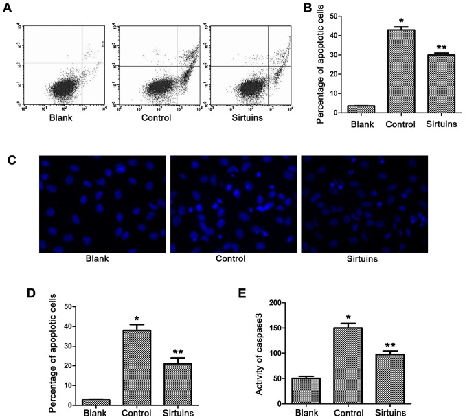
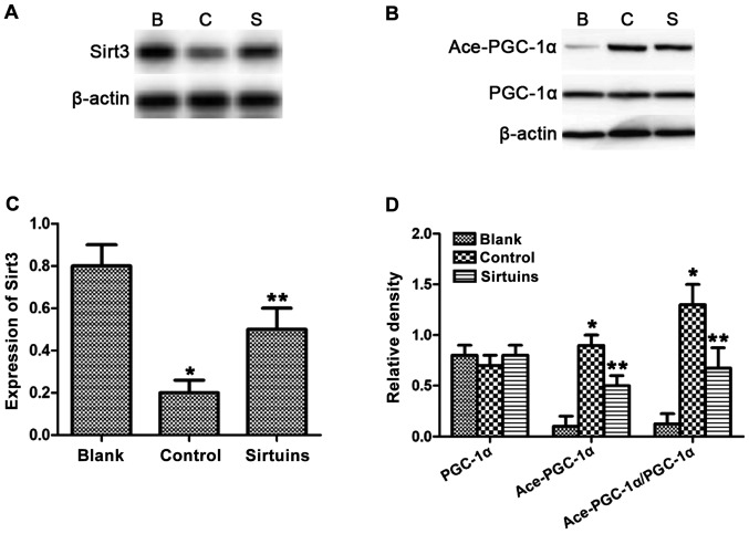
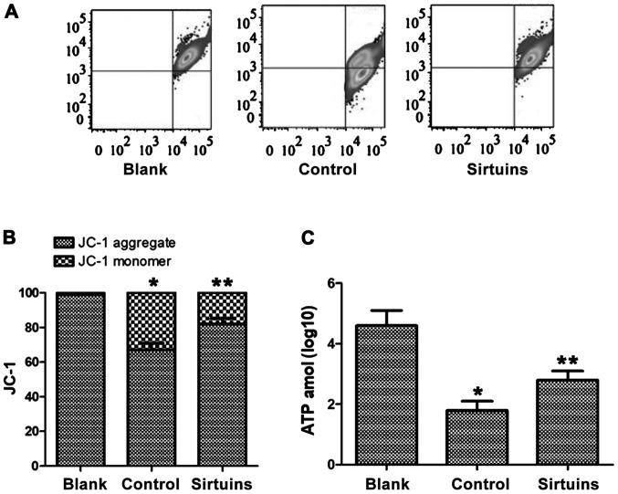
Protective effect of phosphodiesterase 5 (PDE5) inhibitor sildenafil on hypoxic injury of isolated myocardial cells and its mechanism of action were investigated. Myocardial cells of neonatal mice were isolated, cultured and divided into blank, control, and PDE5 inhibitor group. Cells in the control and the PDE5 inhibitor group were treated with hypoxia and serum deprivation for 6 h to simulate the myocardial ischemia process , while those in the PDE5 inhibitor group were treated with 1 µmol/l sildenafil. The cell viability was detected via Cell Counting kit-8 (CCK-8), the cytotoxicity was detected via lactate dehydrogenase release assay, and the apoptosis level was detected via flow cytometry, Hoechst staining and caspase-3 activity assay. Moreover, changes in mitochondrial membrane potential of myocardial cells were detected via JC-1 staining and flow cytometry, fluorescein adenosine triphosphate (ATP) assay kit was used to detect the production of ATP in myocardial cells, and reverse transcription-polymerase chain reaction (RT-PCR) was used to detect the level of Sirt3 messenger ribonucleic acid (mRNA) in myocardial cells. Finally, the expression and changes of Sirt3, peroxisome proliferator-activated receptor γ coactivator-1α (PGC-1α) and acetylated PGC-1α were detected via western blot analysis. After hypoxia treatment, the cell viability was decreased, the cytotoxic effect was enhanced, the percentage of apoptosis was increased, the activity of apoptosis-related protein was increased, the mitochondrial membrane potential was decreased, the production of ATP was reduced, the expression of Sirt3 was decreased, and the acetylation level of PGC-1α was increased. However, after pretreatment with sildenafil, the damage to membrane potential of myocardial cells was significantly alleviated, and the production of ATP was increased. At the same time, myocardial cell apoptosis was decreased, Sirt3 expression was increased and PGC-1α acetylation was decreased. PDE5 inhibitor inhibits apoptosis of hypoxic myocardial cells through protecting mitochondrial function.
研究了磷酸二酯酶5(PDE5)抑制剂西地那非对离体心肌细胞缺氧损伤的保护作用及其作用机制。分离培养新生小鼠心肌细胞,分为空白组、对照组和PDE5抑制剂组。对照组和PDE5抑制剂组细胞进行缺氧缺血清处理6 h以模拟心肌缺血过程,PDE5抑制剂组细胞用1 μmol/L西地那非处理。通过细胞计数试剂盒-8(CCK-8)检测细胞活力,通过乳酸脱氢酶释放试验检测细胞毒性,通过流式细胞术、Hoechst染色和caspase-3活性试验检测细胞凋亡水平。此外,通过JC-1染色和流式细胞术检测心肌细胞线粒体膜电位的变化,用荧光素三磷酸腺苷(ATP)检测试剂盒检测心肌细胞中ATP的产生,用逆转录-聚合酶链反应(RT-PCR)检测心肌细胞中沉默信息调节因子3(Sirt3)信使核糖核酸(mRNA)水平。最后,通过蛋白质免疫印迹分析检测Sirt3、过氧化物酶体增殖物激活受体γ共激活因子-1α(PGC-1α)和乙酰化PGC-1α的表达及变化。缺氧处理后,细胞活力降低,细胞毒性作用增强,凋亡百分比增加,凋亡相关蛋白活性增加,线粒体膜电位降低,ATP产生减少,Sirt3表达降低,PGC-1α乙酰化水平增加。然而,用西地那非预处理后,心肌细胞膜电位损伤明显减轻,ATP产生增加。同时,心肌细胞凋亡减少,Sirt3表达增加,PGC-1α乙酰化减少。PDE5抑制剂通过保护线粒体功能抑制缺氧心肌细胞凋亡。