Multidisciplinary Cardiovascular Research Centre & The Division of Biomedical Imaging, Leeds Institute for Cardiovascular and Metabolic Medicine, University of Leeds, Leeds, UK.
Cognitive Science Research Unit, University of Westminster, London, UK.
BMJ Open. 2019 Jan 21;9(1):e022329. doi: 10.1136/bmjopen-2018-022329.
To compare the incidence of silent cerebral infarction and impact on cognitive function following transcatheter aortic valve implantation (TAVI) with the first-generation CoreValve (Medtronic, Minneapolis, Minnesota, USA) and second-generation Lotus valve (Boston Scientific, Natick Massachusetts, USA).
A prospective observational study comprising a 1.5 T cerebral MRI scan, performed preoperatively and immediately following TAVI, and neurocognitive assessments performed at baseline, 30 days and 1 year follow-up.
University hospitals of Leeds and Leicester, UK.
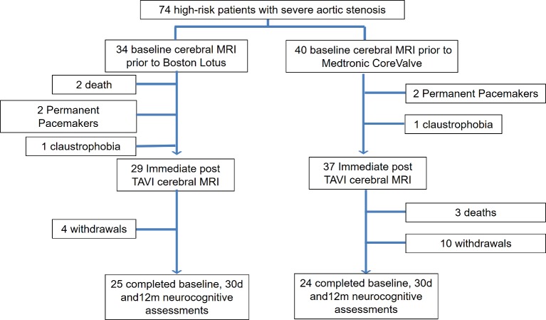
66 (80.6±8.0 years, 47% male) patients with high-risk severe symptomatic aortic stenosis recruited between April 2012 and May 2015.
Incidence of new cerebral microinfarction and objective decline in neurocognitive performance.
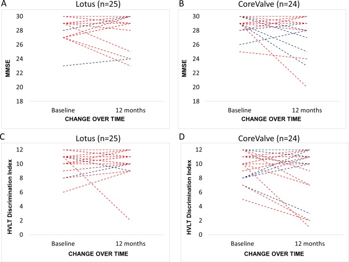
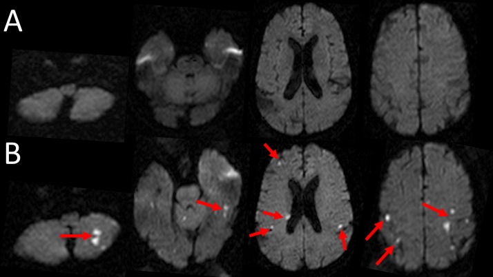
All underwent cerebral MRI at baseline and immediately following TAVI, and 49 (25 Lotus, 24 CoreValve) completed neurocognitive assessments at baseline, 30 days and 1 year. There was a significantly greater incidence of new cerebral microinfarction observed following the Lotus TAVI (23 (79%) vs 22 (59%), p=0.025) with a greater number of new infarcts per patient (median 3.5 (IQR 7.0) vs 2.0 (IQR 3.0), p=0.002). The mean volume of infarcted cerebral tissue per patient was equivalent following the two prostheses (p=0.166). More patients suffered new anterior (14 (48%) vs 2 (5%), p=0.001) and vertebrobasilar (15 (52%) vs 7 (19%), p=0.005) lesions following Lotus. Lotus was associated with a decline in verbal memory and psychomotor speed at 30 days. However, performance longitudinally at 1 year was preserved in all neurocognitive domains.
There was a higher incidence of silent cerebral microinfarction and a greater number of lesions per patient following Lotus compared with CoreValve. However, there was no objective decline in neurocognitive function discernible at 1 year following TAVI with either prosthesis.
比较经导管主动脉瓣植入术(TAVI)后第一代 CoreValve(美敦力,明尼苏达州明尼阿波利斯市)和第二代 Lotus 瓣膜(波士顿科学,马萨诸塞州纳蒂克市)导致的无症状性脑梗死发生率及其对认知功能的影响。
前瞻性观察性研究,包括术前和 TAVI 后即刻行 1.5T 脑 MRI 扫描,并在基线、术后 30 天和 1 年时进行神经认知评估。
英国利兹大学医院和莱斯特大学医院。
2012 年 4 月至 2015 年 5 月期间,共招募了 66 名(80.6±8.0 岁,47%为男性)高危严重症状性主动脉瓣狭窄患者。
新发脑微梗死的发生率和神经认知功能的客观下降。
所有患者均在基线和 TAVI 后即刻行脑 MRI 检查,其中 49 名(25 名 Lotus,24 名 CoreValve)在基线、术后 30 天和 1 年时完成了神经认知评估。Lotus TAVI 后新发脑微梗死的发生率明显更高(23 例[79%]比 22 例[59%],p=0.025),且每位患者新发梗死灶的数量更多(中位数 3.5(IQR 7.0)比 2.0(IQR 3.0),p=0.002)。两种瓣膜术后患者脑梗死组织的平均体积相当(p=0.166)。Lotus 组更多患者新发前循环(14 例[48%]比 2 例[5%],p=0.001)和后循环(15 例[52%]比 7 例[19%],p=0.005)病变。Lotus 与术后 30 天的言语记忆和精神运动速度下降有关。然而,在 TAVI 后 1 年时,所有神经认知领域的功能均保持稳定。
与 CoreValve 相比,Lotus 术后无症状性脑微梗死的发生率更高,每位患者的梗死灶数量更多。然而,在 TAVI 后 1 年,两种瓣膜均未发现神经认知功能的客观下降。