Miyazaki Kenichi, Lisman John E, Ross William N
Department of Physiology, New York Medical College, Valhalla, NY, United States.
Marine Biological Laboratory, Woods Hole, MA, United States.
Front Cell Neurosci. 2019 Jan 8;12:514. doi: 10.3389/fncel.2018.00514. eCollection 2018.
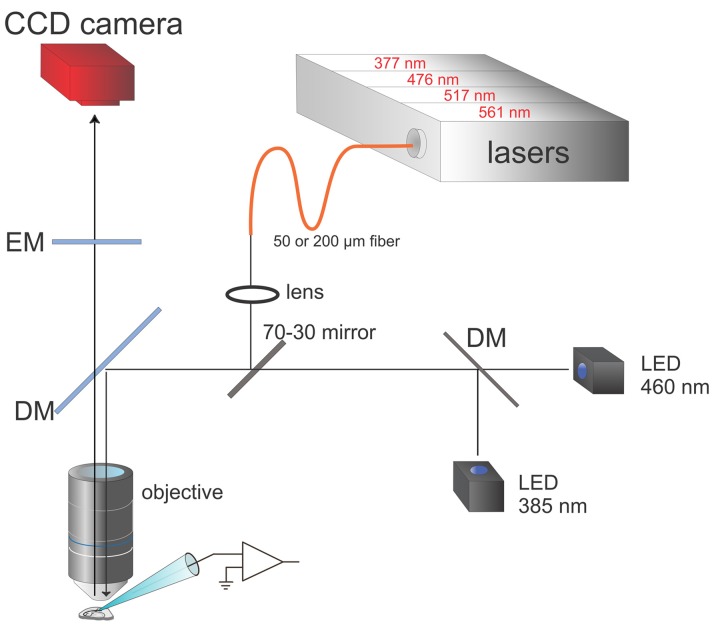
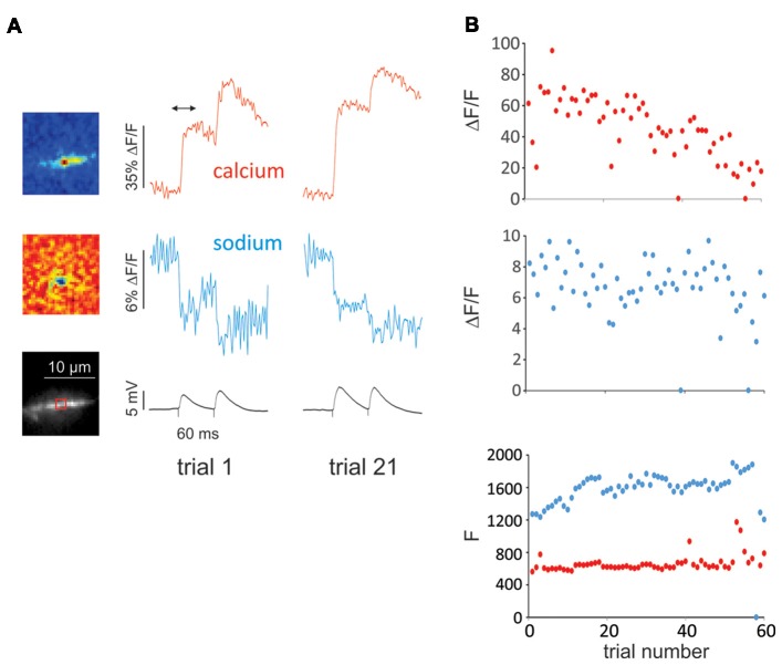
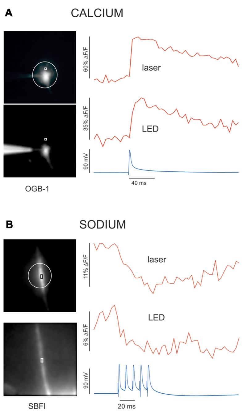
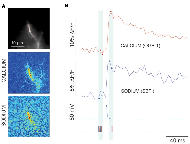
High speed imaging of ion concentration changes in neurons is an important and growing tool for neuroscientists. We previously developed a system for simultaneously measuring sodium and calcium changes in small compartments in neurons (Miyazaki and Ross, 2015). We used this technique to analyze the dynamics of these ions in individual pyramidal neuron dendritic spines (Miyazaki and Ross, 2017). This system is based on high speed multiplexing of light emitting diodes (LEDs) and classic organic indicators. To improve this system we made additional changes, primarily incorporating lasers in addition to the LEDs, more sophisticated imaging protocols, and the use of newer sodium and calcium indicators. This new system generates signals with higher signal to noise ratio (S/N), less background fluorescence, and less photodynamic damage. In addition, by using longer wavelength indicators instead of indicators sensitive in the UV range, it allows for the incorporation of focal uncaging along with simultaneous imaging, which should extend the range of experiments.
对神经元中离子浓度变化进行高速成像,对于神经科学家来说是一种重要且不断发展的工具。我们之前开发了一个系统,用于同时测量神经元小区域内的钠和钙变化(宫崎和罗斯,2015年)。我们使用该技术分析了单个锥体神经元树突棘中这些离子的动态(宫崎和罗斯,2017年)。该系统基于发光二极管(LED)的高速复用和经典有机指示剂。为了改进该系统,我们进行了更多更改,主要是除了LED之外还加入了激光、更复杂的成像方案以及使用更新的钠和钙指示剂。这个新系统产生的信号具有更高的信噪比(S/N)、更少的背景荧光和更少的光动力损伤。此外,通过使用波长更长的指示剂而非对紫外线范围敏感的指示剂,它允许在同时成像的同时结合焦点光解笼锁,这应该会扩展实验范围。