• 文献检索
  • 文档翻译
  • 深度研究
  • 学术资讯
  • Suppr Zotero 插件Zotero 插件
  • 邀请有礼
  • 套餐&价格
  • 历史记录
应用&插件
Suppr Zotero 插件Zotero 插件浏览器插件Mac 客户端Windows 客户端微信小程序
定价
高级版会员购买积分包购买API积分包
服务
文献检索文档翻译深度研究API 文档MCP 服务
关于我们
关于 Suppr公司介绍联系我们用户协议隐私条款
关注我们

Suppr 超能文献

核心技术专利:CN118964589B侵权必究
粤ICP备2023148730 号-1Suppr @ 2026

文献检索

告别复杂PubMed语法,用中文像聊天一样搜索,搜遍4000万医学文献。AI智能推荐,让科研检索更轻松。

立即免费搜索

文件翻译

保留排版,准确专业,支持PDF/Word/PPT等文件格式,支持 12+语言互译。

免费翻译文档

深度研究

AI帮你快速写综述,25分钟生成高质量综述,智能提取关键信息,辅助科研写作。

立即免费体验

现代腕足动物壳中方解石纤维的形成。

Calcite fibre formation in modern brachiopod shells.

机构信息

Department of Earth and Environmental Sciences, LMU, 80333, München, Germany.

Central Facility for Electron Microscopy, University of Ulm, 89069, Ulm, Germany.

出版信息

Sci Rep. 2019 Jan 24;9(1):598. doi: 10.1038/s41598-018-36959-z.

DOI:10.1038/s41598-018-36959-z
PMID:30679565
原文链接:https://pmc.ncbi.nlm.nih.gov/articles/PMC6345923/
Abstract

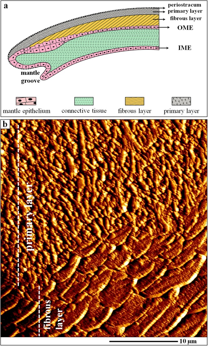
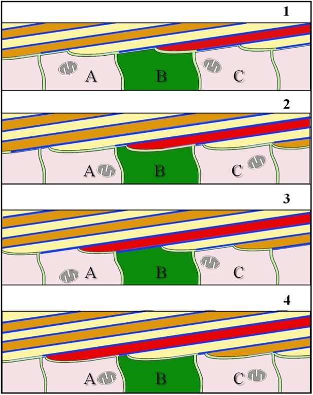
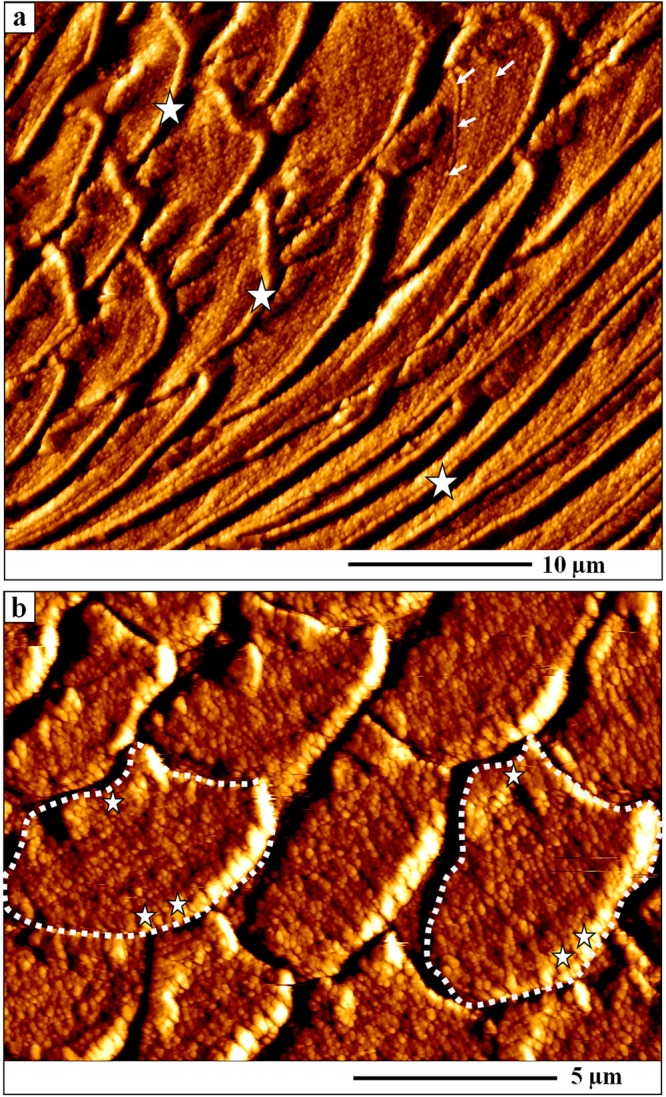
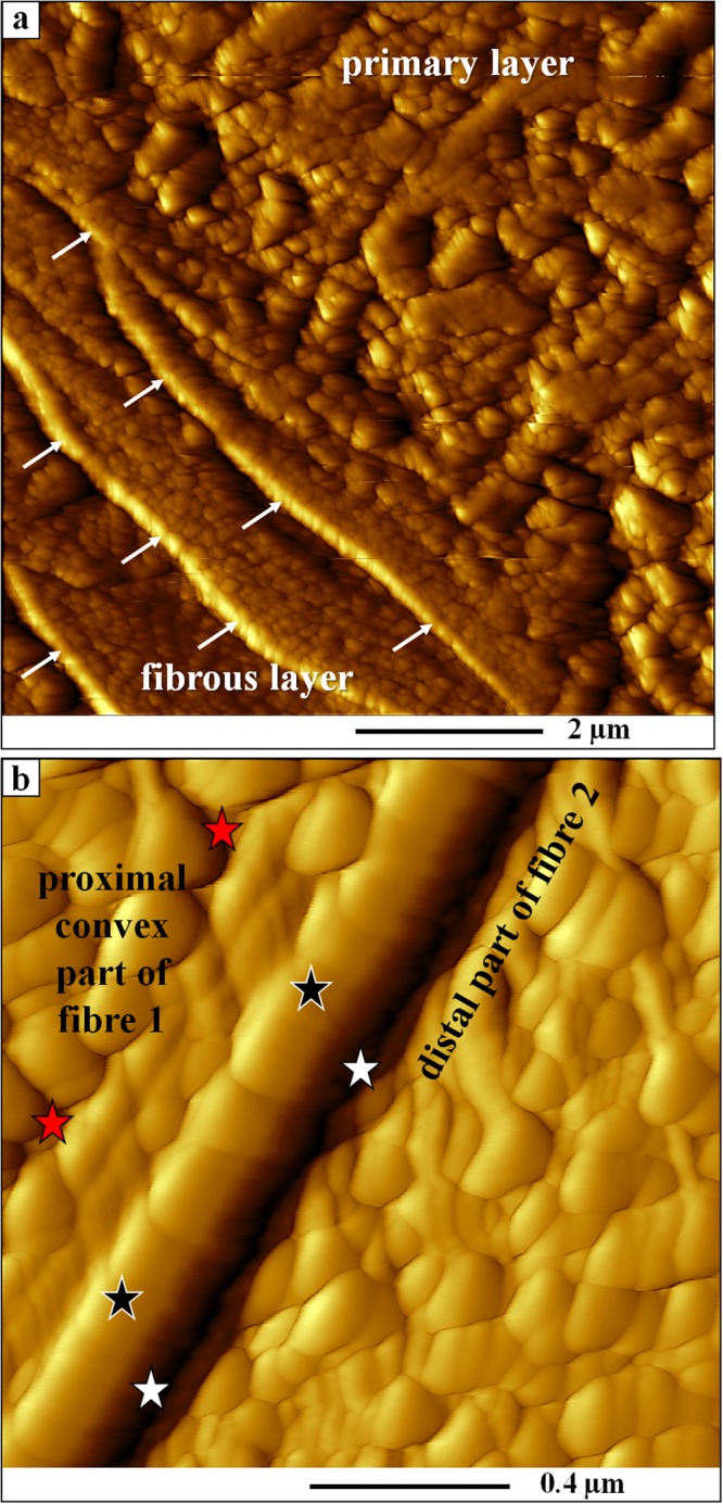
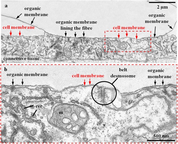
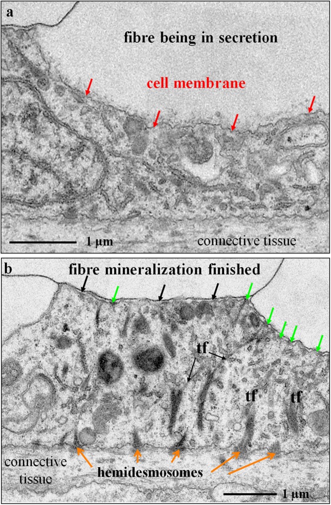
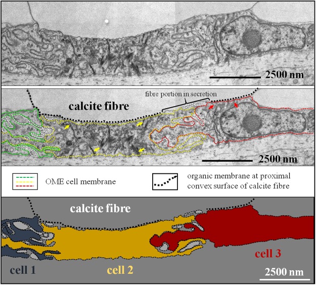
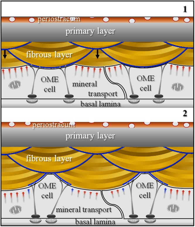
The fibrous calcite layer of modern brachiopod shells is a hybrid composite material and forms a substantial part of the hard tissue. We investigated how cells of the outer mantle epithelium (OME) secrete calcite material and generate the characteristic fibre morphology and composite microstructure of the shell. We employed AFM, FE-SEM, and TEM imaging of embedded/etched, chemically fixed/decalcified and high-pressure frozen/freeze substituted samples. Calcite fibres are secreted by outer mantle epithelium (OME) cells. Biometric analysis of TEM micrographs indicates that about 50% of these cells are attached via hemidesmosomes to an extracellular organic membrane present at the proximal, convex surface of the fibres. At these sites, mineral secretion is not active. Instead, ion transport from OME cells to developing fibres occurs at regions of closest contact between cells and fibres, however only at sites where the extracellular membrane at the proximal fibre surface is not developed yet. Fibre formation requires the cooperation of several adjacent OME cells. It is a spatially and temporally changing process comprising of detachment of OME cells from the extracellular organic membrane, mineral secretion at detachment sites, termination of secretion with formation of the extracellular organic membrane, and attachment of cells via hemidesmosomes to this membrane.

摘要

现代腕足动物壳的纤维方解石层是一种混合复合材料,构成了硬组织的重要部分。我们研究了外套膜上皮(OME)细胞如何分泌方解石物质并产生壳的特征纤维形态和复合微观结构。我们采用 AFM、FE-SEM 和 TEM 成像技术,对嵌入式/蚀刻、化学固定/脱钙和高压冷冻/冷冻替代的样本进行了研究。方解石纤维由外套膜上皮(OME)细胞分泌。TEM 显微照片的生物计量分析表明,这些细胞中约有 50%通过半桥粒附着在纤维近端凸面存在的细胞外有机膜上。在这些部位,矿化分泌不活跃。相反,离子从 OME 细胞向发育中的纤维的运输发生在细胞和纤维之间的最紧密接触区域,但仅在细胞外膜在近端纤维表面尚未发育的部位发生。纤维的形成需要几个相邻的 OME 细胞的合作。这是一个空间和时间上不断变化的过程,包括 OME 细胞从细胞外有机膜上的分离、分离部位的矿化分泌、分泌终止和形成细胞外有机膜,以及通过半桥粒附着到该膜上的细胞。

https://cdn.ncbi.nlm.nih.gov/pmc/blobs/80f5/6345923/eb150ccb4e12/41598_2018_36959_Fig9_HTML.jpg
https://cdn.ncbi.nlm.nih.gov/pmc/blobs/80f5/6345923/708bcda9c27a/41598_2018_36959_Fig1_HTML.jpg
https://cdn.ncbi.nlm.nih.gov/pmc/blobs/80f5/6345923/0e552f4365b2/41598_2018_36959_Fig2_HTML.jpg
https://cdn.ncbi.nlm.nih.gov/pmc/blobs/80f5/6345923/a74d33e74460/41598_2018_36959_Fig3_HTML.jpg
https://cdn.ncbi.nlm.nih.gov/pmc/blobs/80f5/6345923/f9dfbdef84da/41598_2018_36959_Fig4_HTML.jpg
https://cdn.ncbi.nlm.nih.gov/pmc/blobs/80f5/6345923/13f9fc8cfa5b/41598_2018_36959_Fig5_HTML.jpg
https://cdn.ncbi.nlm.nih.gov/pmc/blobs/80f5/6345923/897d89e15443/41598_2018_36959_Fig6_HTML.jpg
https://cdn.ncbi.nlm.nih.gov/pmc/blobs/80f5/6345923/1cff1cd3523f/41598_2018_36959_Fig7_HTML.jpg
https://cdn.ncbi.nlm.nih.gov/pmc/blobs/80f5/6345923/7033a3a6a6cd/41598_2018_36959_Fig8_HTML.jpg
https://cdn.ncbi.nlm.nih.gov/pmc/blobs/80f5/6345923/eb150ccb4e12/41598_2018_36959_Fig9_HTML.jpg

相似文献

1
Calcite fibre formation in modern brachiopod shells.现代腕足动物壳中方解石纤维的形成。
Sci Rep. 2019 Jan 24;9(1):598. doi: 10.1038/s41598-018-36959-z.
2
Terebratulide brachiopod shell biomineralization by mantle epithelial cells.腕足动物瓣鳃纲贝壳生物矿化作用由套膜上皮细胞完成。
J Struct Biol. 2019 Aug 1;207(2):136-157. doi: 10.1016/j.jsb.2019.05.002. Epub 2019 May 6.
3
Biogenic calcite granules--are brachiopods different?生物成因方解石颗粒——腕足动物有何不同?
Micron. 2013 Jan;44:395-403. doi: 10.1016/j.micron.2012.09.005. Epub 2012 Sep 16.
4
Multiscale structure of calcite fibres of the shell of the brachiopod Terebratulina retusa.腕足动物钝圆穿孔贝类贝壳中方解石纤维的多尺度结构
J Struct Biol. 2008 Oct;164(1):96-100. doi: 10.1016/j.jsb.2008.06.010. Epub 2008 Jun 27.
5
Mapping of recent brachiopod microstructure: A tool for environmental studies.近期腕足动物微观结构的绘图:环境研究的工具。
J Struct Biol. 2018 Mar;201(3):221-236. doi: 10.1016/j.jsb.2017.11.011. Epub 2017 Nov 23.
6
Biological strategy for the fabrication of highly ordered aragonite helices: the microstructure of the cavolinioidean gastropods.制造高度有序文石螺旋的生物学策略:卡沃利尼腹足纲动物的微观结构。
Sci Rep. 2016 May 16;6:25989. doi: 10.1038/srep25989.
7
The structural, compositional and mechanical features of the calcite shell of the barnacle Tetraclita rufotincta.藤壶 Tetraclita rufotincta 碳酸钙贝壳的结构、组成和力学特性。
J Struct Biol. 2011 Sep;175(3):311-8. doi: 10.1016/j.jsb.2011.04.014. Epub 2011 Apr 28.
8
Initial formation of calcite crystals in the thin prismatic layer with the periostracum of Pinctada fucata.在珍珠贝的薄棱柱层和外皮的初始碳酸钙晶体形成。
Micron. 2013 Feb;45:136-9. doi: 10.1016/j.micron.2012.10.010. Epub 2012 Oct 29.
9
The Magellania venosa Biomineralizing Proteome: A Window into Brachiopod Shell Evolution.麦哲伦脉纹贝生物矿化蛋白质组:了解腕足动物贝壳演化的窗口。
Genome Biol Evol. 2015 Apr 24;7(5):1349-62. doi: 10.1093/gbe/evv074.
10
Orientation patterns of aragonitic crossed-lamellar, fibrous prismatic and myostracal microstructures of modern Glycymeris shells.现代甘氨酸贝壳的文石交叉层、纤维状棱柱和肌质微结构的取向模式。
J Struct Biol. 2020 Dec 1;212(3):107653. doi: 10.1016/j.jsb.2020.107653. Epub 2020 Oct 23.

引用本文的文献

1
Statistical analysis of EBSD data confirms pronounced classical and non-classical pervasive crystallographic twinning in rotaliid foraminiferal calcite.电子背散射衍射(EBSD)数据的统计分析证实,在轮虫类有孔虫方解石中存在明显的经典和非经典普遍晶体孪生现象。
Sci Rep. 2025 Apr 28;15(1):14852. doi: 10.1038/s41598-025-92636-y.
2
Skeletal microstructures of cheilostome bryozoans (phylum Bryozoa, class Gymnolaemata): crystallography and secretion patterns.唇口目苔藓虫(苔藓虫纲,裸唇亚纲)的骨骼微结构:晶体学与分泌模式
Mar Life Sci Technol. 2024 Jun 7;6(3):405-424. doi: 10.1007/s42995-024-00233-1. eCollection 2024 Aug.
3
Biomineral crystallographic preferred orientation in Solenogastres molluscs (Aplacophora) is controlled by organic templating.

本文引用的文献

1
Microstructural data of six recent brachiopod species: SEM, EBSD, morphometric and statistical analyses.六种现代腕足动物的微观结构数据:扫描电子显微镜、电子背散射衍射、形态测量与统计分析
Data Brief. 2018 Mar 6;18:300-318. doi: 10.1016/j.dib.2018.02.071. eCollection 2018 Jun.
2
Mapping of recent brachiopod microstructure: A tool for environmental studies.近期腕足动物微观结构的绘图:环境研究的工具。
J Struct Biol. 2018 Mar;201(3):221-236. doi: 10.1016/j.jsb.2017.11.011. Epub 2017 Nov 23.
3
Organic membranes determine the pattern of the columnar prismatic layer of mollusc shells.
棘皮动物门软体动物(无板纲)的生物矿化晶体结晶各向异性受有机模板控制。
Sci Rep. 2024 May 5;14(1):10309. doi: 10.1038/s41598-024-57754-z.
4
Correlative chemical and elemental nano-imaging of morphology and disorder at the nacre-prismatic region interface in Pinctada margaritifera.马氏珠母贝珍珠层-棱柱层区域界面形态与无序性的相关化学和元素纳米成像
Sci Rep. 2023 Dec 1;13(1):21258. doi: 10.1038/s41598-023-47446-5.
5
Cell type and gene regulatory network approaches in the evolution of spiralian biomineralisation.螺旋动物生物矿化进化中的细胞类型和基因调控网络方法。
Brief Funct Genomics. 2023 Nov 17;22(6):509-516. doi: 10.1093/bfgp/elad033.
6
The argonaut constructs its shell via physical self-organization and coordinated cell sensorial activity.船蛸通过物理自组织和协调的细胞感官活动构建其外壳。
iScience. 2021 Oct 15;24(11):103288. doi: 10.1016/j.isci.2021.103288. eCollection 2021 Nov 19.
7
Origin of the biphase nature and surface roughness of biogenic calcite secreted by the giant barnacle Austromegabalanus psittacus.巨型藤壶 Austromegabalanus psittacus 分泌的生物成因方解石的双相性质和表面粗糙度的起源。
Sci Rep. 2020 Oct 8;10(1):16784. doi: 10.1038/s41598-020-73804-8.
8
Foamy oysters: vesicular microstructure production in the Gryphaeidae via emulsification.泡沫贻贝:通过乳化作用在 Gryphaeidae 中产生泡沫状微观结构。
J R Soc Interface. 2020 Sep;17(170):20200505. doi: 10.1098/rsif.2020.0505. Epub 2020 Sep 30.
9
Microstructure and crystallography of the wall plates of the giant barnacle : a material organized by crystal growth.巨型藤壶壁板的微观结构和晶体学:一种由晶体生长组织的材料。
J R Soc Interface. 2020 Mar;17(164):20190743. doi: 10.1098/rsif.2019.0743. Epub 2020 Mar 4.
10
Mechanics unlocks the morphogenetic puzzle of interlocking bivalved shells.力学解开了联锁双壳贝类形态发生的谜题。
Proc Natl Acad Sci U S A. 2020 Jan 7;117(1):43-51. doi: 10.1073/pnas.1916520116. Epub 2019 Dec 16.
有机膜决定了软体动物贝壳柱状棱柱层的形态。
Proc Biol Sci. 2016 May 11;283(1830). doi: 10.1098/rspb.2016.0032.
4
Structural Design Elements in Biological Materials: Application to Bioinspiration.生物材料的结构设计元素:仿生学应用。
Adv Mater. 2015 Oct 7;27(37):5455-76. doi: 10.1002/adma.201502403. Epub 2015 Aug 25.
5
Bioinspired structural materials.仿生结构材料。
Nat Mater. 2015 Jan;14(1):23-36. doi: 10.1038/nmat4089. Epub 2014 Oct 26.
6
Tailored order: the mesocrystalline nature of sea urchin teeth.定制订单:海胆牙齿的介观晶体性质。
Acta Biomater. 2014 Sep;10(9):3885-98. doi: 10.1016/j.actbio.2014.06.012. Epub 2014 Jun 14.
7
Surviving the surf: the tribomechanical properties of the periostracum of Mytilus sp.在海浪中幸存:贻贝属的壳皮的摩擦力学性能
Acta Biomater. 2014 Sep;10(9):3978-85. doi: 10.1016/j.actbio.2014.05.014. Epub 2014 May 23.
8
Bioinspired materials that self-shape through programmed microstructures.通过程序控制微观结构实现自形状的仿生材料。
Soft Matter. 2014 Mar 7;10(9):1284-94. doi: 10.1039/c3sm51883c.
9
Biological control of crystallographic architecture: hierarchy and co-alignment parameters.生物控制结晶结构:层次和共取向参数。
Acta Biomater. 2014 Sep;10(9):3866-74. doi: 10.1016/j.actbio.2014.02.039. Epub 2014 Feb 28.
10
The quest for stiff, strong and tough hybrid materials: an exhaustive exploration.追求坚硬、强韧的混合材料:全面探索。
J R Soc Interface. 2013 Sep 25;10(89):20130711. doi: 10.1098/rsif.2013.0711. Print 2013 Dec 6.