• 文献检索
  • 文档翻译
  • 深度研究
  • 学术资讯
  • Suppr Zotero 插件Zotero 插件
  • 邀请有礼
  • 套餐&价格
  • 历史记录
应用&插件
Suppr Zotero 插件Zotero 插件浏览器插件Mac 客户端Windows 客户端微信小程序
定价
高级版会员购买积分包购买API积分包
服务
文献检索文档翻译深度研究API 文档MCP 服务
关于我们
关于 Suppr公司介绍联系我们用户协议隐私条款
关注我们

Suppr 超能文献

核心技术专利:CN118964589B侵权必究
粤ICP备2023148730 号-1Suppr @ 2026

文献检索

告别复杂PubMed语法,用中文像聊天一样搜索,搜遍4000万医学文献。AI智能推荐,让科研检索更轻松。

立即免费搜索

文件翻译

保留排版,准确专业,支持PDF/Word/PPT等文件格式,支持 12+语言互译。

免费翻译文档

深度研究

AI帮你快速写综述,25分钟生成高质量综述,智能提取关键信息,辅助科研写作。

立即免费体验

慢性神经退行性病变通过 STING 诱导 I 型干扰素合成,塑造小胶质细胞表型并加速疾病进展。

Chronic neurodegeneration induces type I interferon synthesis via STING, shaping microglial phenotype and accelerating disease progression.

机构信息

School of Biochemistry and Immunology, Trinity College Institute of Neuroscience & Trinity Biomedical Sciences Institute, Trinity College Dublin, Dublin, Republic of Ireland.

Department of Medicine, Division of Pulmonary, Critical Care and Sleep Medicine, University of Florida, Gainesville, Florida.

出版信息

Glia. 2019 Jul;67(7):1254-1276. doi: 10.1002/glia.23592. Epub 2019 Jan 25.

DOI:10.1002/glia.23592
PMID:30680794
原文链接:https://pmc.ncbi.nlm.nih.gov/articles/PMC6520218/
Abstract

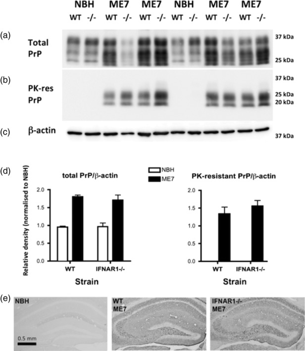
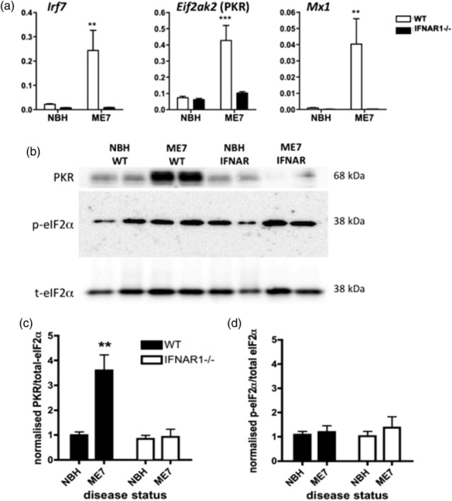
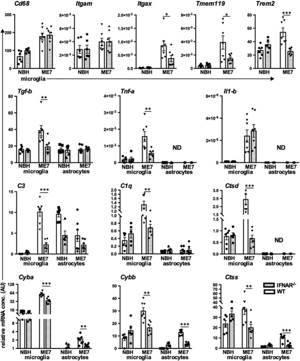
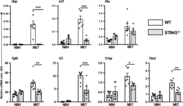
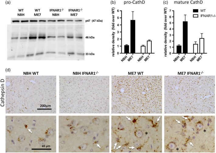
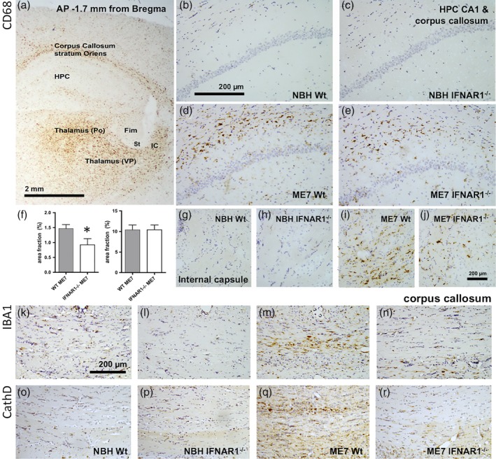
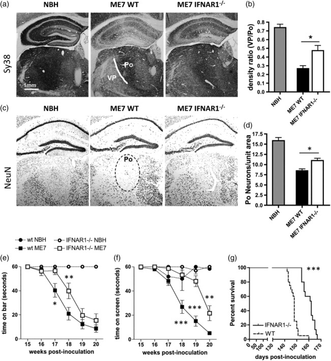
Type I interferons (IFN-I) are the principal antiviral molecules of the innate immune system and can be made by most cell types, including central nervous system cells. IFN-I has been implicated in neuroinflammation during neurodegeneration, but its mechanism of induction and its consequences remain unclear. In the current study, we assessed expression of IFN-I in murine prion disease (ME7) and examined the contribution of the IFN-I receptor IFNAR1 to disease progression. The data indicate a robust IFNβ response, specifically in microglia, with evidence of IFN-dependent genes in both microglia and astrocytes. This IFN-I response was absent in stimulator of interferon genes (STING ) mice. Microglia showed increased numbers and activated morphology independent of genotype, but transcriptional signatures indicated an IFNAR1-dependent neuroinflammatory phenotype. Isolation of microglia and astrocytes demonstrated disease-associated microglial induction of Tnfα, Tgfb1, and of phagolysosomal system transcripts including those for cathepsins, Cd68, C1qa, C3, and Trem2, which were diminished in IFNAR1 and STING deficient mice. Microglial increases in activated cathepsin D, and CD68 were significantly reduced in IFNAR1 mice, particularly in white matter, and increases in COX-1 expression, and prostaglandin synthesis were significantly mitigated. Disease progressed more slowly in IFNAR1 mice, with diminished synaptic and neuronal loss and delayed onset of neurological signs and death but without effect on proteinase K-resistant PrP levels. Therefore, STING-dependent IFN-I influences microglial phenotype and influences neurodegenerative progression despite occurring secondary to initial degenerative changes. These data expand our mechanistic understanding of IFN-I induction and its impact on microglial function during chronic neurodegeneration.

摘要

I 型干扰素 (IFN-I) 是先天免疫系统的主要抗病毒分子,可由包括中枢神经系统细胞在内的大多数细胞类型产生。IFN-I 已被牵连到神经退行性变中的神经炎症中,但诱导机制及其后果仍不清楚。在当前的研究中,我们评估了 IFN-I 在鼠朊病毒病 (ME7) 中的表达,并研究了 IFN-I 受体 IFNAR1 对疾病进展的贡献。数据表明存在强烈的 IFNβ 反应,特别是在小胶质细胞中,并且在小胶质细胞和星形胶质细胞中都有 IFN 依赖性基因的证据。这种 IFN-I 反应在干扰素基因刺激因子 (STING) 小鼠中不存在。小胶质细胞的数量增加且形态激活,但转录特征表明存在依赖 IFNAR1 的神经炎症表型。小胶质细胞和星形胶质细胞的分离表明,疾病相关的小胶质细胞诱导了 TNFα、Tgfb1 和吞噬溶酶体系统转录物,包括组织蛋白酶、CD68、C1qa、C3 和 Trem2 的转录物,这些在 IFNAR1 和 STING 缺陷型小鼠中减少。IFNAR1 小鼠中疾病相关的小胶质细胞激活的组织蛋白酶 D 和 CD68 显著减少,特别是在白质中,COX-1 表达和前列腺素合成的增加显著减轻。IFNAR1 小鼠的疾病进展更缓慢,突触和神经元丢失减少,神经症状和死亡的发作延迟,但对蛋白酶 K 抗性 PrP 水平没有影响。因此,STING 依赖性 IFN-I 影响小胶质细胞表型,并影响神经退行性进展,尽管发生在初始退行性变化之后。这些数据扩展了我们对 IFN-I 诱导及其在慢性神经退行性变中小胶质细胞功能中的作用的机制理解。

https://cdn.ncbi.nlm.nih.gov/pmc/blobs/c590/6563482/a6945de0db3e/GLIA-67-1254-g011.jpg
https://cdn.ncbi.nlm.nih.gov/pmc/blobs/c590/6563482/e69d7d4195ef/GLIA-67-1254-g001.jpg
https://cdn.ncbi.nlm.nih.gov/pmc/blobs/c590/6563482/04087d0c2397/GLIA-67-1254-g002.jpg
https://cdn.ncbi.nlm.nih.gov/pmc/blobs/c590/6563482/2f9ee2fd89b0/GLIA-67-1254-g003.jpg
https://cdn.ncbi.nlm.nih.gov/pmc/blobs/c590/6563482/c797399f2d4b/GLIA-67-1254-g004.jpg
https://cdn.ncbi.nlm.nih.gov/pmc/blobs/c590/6563482/27b7b1f49c18/GLIA-67-1254-g005.jpg
https://cdn.ncbi.nlm.nih.gov/pmc/blobs/c590/6563482/9131e0cbd9a7/GLIA-67-1254-g006.jpg
https://cdn.ncbi.nlm.nih.gov/pmc/blobs/c590/6563482/2ba80da0f87f/GLIA-67-1254-g007.jpg
https://cdn.ncbi.nlm.nih.gov/pmc/blobs/c590/6563482/777f8ccfc6e2/GLIA-67-1254-g008.jpg
https://cdn.ncbi.nlm.nih.gov/pmc/blobs/c590/6563482/42e6a68923e2/GLIA-67-1254-g009.jpg
https://cdn.ncbi.nlm.nih.gov/pmc/blobs/c590/6563482/845eb3bbf847/GLIA-67-1254-g010.jpg
https://cdn.ncbi.nlm.nih.gov/pmc/blobs/c590/6563482/a6945de0db3e/GLIA-67-1254-g011.jpg

相似文献

1
Chronic neurodegeneration induces type I interferon synthesis via STING, shaping microglial phenotype and accelerating disease progression.慢性神经退行性病变通过 STING 诱导 I 型干扰素合成,塑造小胶质细胞表型并加速疾病进展。
Glia. 2019 Jul;67(7):1254-1276. doi: 10.1002/glia.23592. Epub 2019 Jan 25.
2
Ischemia/Reperfusion Induces Interferon-Stimulated Gene Expression in Microglia.缺血/再灌注诱导小胶质细胞中干扰素刺激基因的表达。
J Neurosci. 2017 Aug 23;37(34):8292-8308. doi: 10.1523/JNEUROSCI.0725-17.2017. Epub 2017 Jul 26.
3
Microglial responses to peripheral type 1 interferon.小胶质细胞对周围型 I 型干扰素的反应。
J Neuroinflammation. 2020 Nov 12;17(1):340. doi: 10.1186/s12974-020-02003-z.
4
Interferon-β Plays a Detrimental Role in Experimental Traumatic Brain Injury by Enhancing Neuroinflammation That Drives Chronic Neurodegeneration.干扰素-β 通过增强神经炎症促进慢性神经退行性变,在实验性创伤性脑损伤中起有害作用。
J Neurosci. 2020 Mar 11;40(11):2357-2370. doi: 10.1523/JNEUROSCI.2516-19.2020. Epub 2020 Feb 6.
5
DNA sensors are expressed in astrocytes and microglia in vitro and are upregulated during gliosis in neurodegenerative disease.DNA传感器在体外的星形胶质细胞和小胶质细胞中表达,并在神经退行性疾病的胶质增生过程中上调。
Glia. 2015 May;63(5):812-25. doi: 10.1002/glia.22786. Epub 2015 Jan 27.
6
Impaired cortical neuronal homeostasis and cognition after diffuse traumatic brain injury are dependent on microglia and type I interferon responses.弥漫性创伤性脑损伤后皮质神经元内环境稳定和认知功能受损依赖于小胶质细胞和I型干扰素反应。
Glia. 2024 Feb;72(2):300-321. doi: 10.1002/glia.24475. Epub 2023 Nov 8.
7
Positive feedback regulation of type I interferon by the interferon-stimulated gene STING.干扰素刺激基因STING对I型干扰素的正反馈调节
EMBO Rep. 2015 Feb;16(2):202-12. doi: 10.15252/embr.201439366. Epub 2015 Jan 8.
8
Traumatic Brain Injury Induces cGAS Activation and Type I Interferon Signaling in Aged Mice.创伤性脑损伤在老年小鼠中诱导 cGAS 激活和 I 型干扰素信号通路。
Front Immunol. 2021 Aug 24;12:710608. doi: 10.3389/fimmu.2021.710608. eCollection 2021.
9
Accumulation of Cytoplasmic DNA Due to ATM Deficiency Activates the Microglial Viral Response System with Neurotoxic Consequences.由于 ATM 缺陷导致细胞质 DNA 积累,激活了具有神经毒性后果的小胶质细胞病毒反应系统。
J Neurosci. 2019 Aug 7;39(32):6378-6394. doi: 10.1523/JNEUROSCI.0774-19.2019. Epub 2019 Jun 12.
10
Activation of the STING-Dependent Type I Interferon Response Reduces Microglial Reactivity and Neuroinflammation.STING 依赖性 I 型干扰素反应的激活可降低小胶质细胞的反应性和神经炎症。
Neuron. 2017 Dec 20;96(6):1290-1302.e6. doi: 10.1016/j.neuron.2017.11.032.

引用本文的文献

1
Type I interferons regulate nitric oxide production in -activated microglia contributing to neuronal death.I型干扰素调节活化小胶质细胞中一氧化氮的产生,导致神经元死亡。
Front Immunol. 2025 Aug 27;16:1661395. doi: 10.3389/fimmu.2025.1661395. eCollection 2025.
2
Knockout of P2Y12 receptor facilitates neuronal envelopment by reactive microglia and accelerates prion disease.P2Y12受体基因敲除促进反应性小胶质细胞对神经元的包裹并加速朊病毒病。
J Neuroinflammation. 2025 Aug 31;22(1):210. doi: 10.1186/s12974-025-03542-z.
3
Beyond interferons: Non-canonical roles of MITA/STING.

本文引用的文献

1
STING-mediated type-I interferons contribute to the neuroinflammatory process and detrimental effects following traumatic brain injury.STING 介导的 I 型干扰素有助于创伤性脑损伤后的神经炎症过程和不良影响。
J Neuroinflammation. 2018 Nov 21;15(1):323. doi: 10.1186/s12974-018-1354-7.
2
Activation of the STING-Dependent Type I Interferon Response Reduces Microglial Reactivity and Neuroinflammation.STING 依赖性 I 型干扰素反应的激活可降低小胶质细胞的反应性和神经炎症。
Neuron. 2017 Dec 20;96(6):1290-1302.e6. doi: 10.1016/j.neuron.2017.11.032.
3
Temporal Tracking of Microglia Activation in Neurodegeneration at Single-Cell Resolution.
超越干扰素:MITA/STING的非经典作用
Cell Insight. 2025 Jul 24;4(5):100266. doi: 10.1016/j.cellin.2025.100266. eCollection 2025 Oct.
4
The STING Signaling: A Novel Target for Central Nervous System Diseases.STING信号通路:中枢神经系统疾病的新靶点。
Cell Mol Neurobiol. 2025 Apr 7;45(1):33. doi: 10.1007/s10571-025-01550-4.
5
Targeting Ferroptosis in Parkinson's Disease: Mechanisms and Emerging Therapeutic Strategies.帕金森病中铁死亡的靶向治疗:机制与新兴治疗策略
Int J Mol Sci. 2024 Dec 4;25(23):13042. doi: 10.3390/ijms252313042.
6
Supportive care or exhausted neglect: the role of microglia at the end stage of prion disease.支持性护理还是疲惫性忽视:小胶质细胞在朊病毒疾病末期的作用
J Clin Invest. 2024 Dec 2;134(23):e186940. doi: 10.1172/JCI186940.
7
Interleukin-6 deficiency reduces neuroinflammation by inhibiting the STAT3-cGAS-STING pathway in Alzheimer's disease mice.白细胞介素-6 缺乏通过抑制阿尔茨海默病小鼠中的 STAT3-cGAS-STING 通路减少神经炎症。
J Neuroinflammation. 2024 Nov 1;21(1):282. doi: 10.1186/s12974-024-03277-3.
8
Mitochondrial dysfunction, cause or consequence in neurodegenerative diseases?线粒体功能障碍,是神经退行性疾病的病因还是后果?
Bioessays. 2025 Jan;47(1):e2400023. doi: 10.1002/bies.202400023. Epub 2024 Oct 4.
9
Protein kinase R induced by type I interferons is a main regulator of reactive microglia in Zika virus infection.I型干扰素诱导的蛋白激酶R是寨卡病毒感染中反应性小胶质细胞的主要调节因子。
Glia. 2025 Jan;73(1):80-104. doi: 10.1002/glia.24619. Epub 2024 Oct 3.
10
Mitophagy and cGAS-STING crosstalk in neuroinflammation.神经炎症中的线粒体自噬与cGAS-STING相互作用
Acta Pharm Sin B. 2024 Aug;14(8):3327-3361. doi: 10.1016/j.apsb.2024.05.012. Epub 2024 May 13.
单细胞分辨率下神经退行性变中小胶质细胞激活的时间追踪。
Cell Rep. 2017 Oct 10;21(2):366-380. doi: 10.1016/j.celrep.2017.09.039.
4
A Unique Microglia Type Associated with Restricting Development of Alzheimer's Disease.一种与限制阿尔茨海默病发展相关的独特小胶质细胞类型。
Cell. 2017 Jun 15;169(7):1276-1290.e17. doi: 10.1016/j.cell.2017.05.018. Epub 2017 Jun 8.
5
Extracellular histones, cell-free DNA, or nucleosomes: differences in immunostimulation.细胞外组蛋白、游离DNA或核小体:免疫刺激方面的差异
Cell Death Dis. 2016 Dec 8;7(12):e2518. doi: 10.1038/cddis.2016.410.
6
Endoplasmic Reticulum Stress-induced Hepatocellular Death Pathways Mediate Liver Injury and Fibrosis via Stimulator of Interferon Genes.内质网应激诱导的肝细胞死亡途径通过干扰素基因刺激因子介导肝损伤和肝纤维化。
J Biol Chem. 2016 Dec 23;291(52):26794-26805. doi: 10.1074/jbc.M116.736991. Epub 2016 Nov 3.
7
Regulation and function of the cGAS-STING pathway of cytosolic DNA sensing.细胞质 DNA 感应的 cGAS-STING 途径的调控和功能。
Nat Immunol. 2016 Sep 20;17(10):1142-9. doi: 10.1038/ni.3558.
8
Type-1 interferons contribute to the neuroinflammatory response and disease progression of the MPTP mouse model of Parkinson's disease.1型干扰素促成帕金森病MPTP小鼠模型的神经炎症反应和疾病进展。
Glia. 2016 Sep;64(9):1590-604. doi: 10.1002/glia.23028. Epub 2016 Jul 12.
9
Deletion of the type-1 interferon receptor in APPSWE/PS1ΔE9 mice preserves cognitive function and alters glial phenotype.在 APPswe/PS1ΔE9 小鼠中敲除 I 型干扰素受体可保持认知功能并改变神经胶质表型。
Acta Neuropathol Commun. 2016 Jul 11;4(1):72. doi: 10.1186/s40478-016-0341-4.
10
Brain Endothelial- and Epithelial-Specific Interferon Receptor Chain 1 Drives Virus-Induced Sickness Behavior and Cognitive Impairment.脑内皮细胞和上皮细胞特异性干扰素受体链 1 驱动病毒诱导的疾病行为和认知障碍。
Immunity. 2016 Apr 19;44(4):901-12. doi: 10.1016/j.immuni.2016.04.005.