Department of Pharmacology, Nihon University School of Dentistry, Chiyoda-ku, Tokyo 101-8310, Japan.
Division of Oral and Craniomaxillofacial Research, Dental Research Center, Nihon University School of Dentistry, Chiyoda-ku, Tokyo 101-8310, Japan.
eNeuro. 2019 Jan 28;6(1). doi: 10.1523/ENEURO.0462-18.2019. eCollection 2019 Jan-Feb.
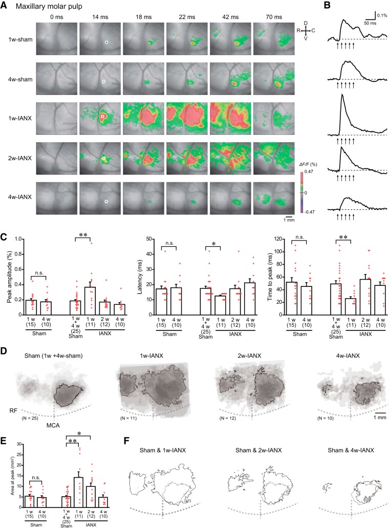
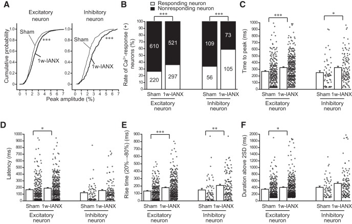
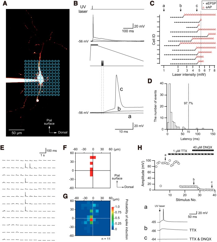
The primary sensory cortex processes competitive sensory inputs. Ablation of these competitive inputs induces neuroplastic changes in local cortical circuits. However, information concerning cortical plasticity induced by a disturbance of competitive nociceptive inputs is limited. Nociceptive information from the maxillary and mandibular molar pulps converges at the border between the ventral secondary somatosensory cortex (S2) and insular oral region (IOR); therefore, S2/IOR is a suitable target for examining the cortical changes induced by a disturbance of noxious inputs, which often causes neuropathic pain and allodynia. We focused on the plastic changes in S2/IOR excitation in a model of rats subjected to inferior alveolar nerve transection (IANX). Our optical imaging using a voltage-sensitive dye (VSD) revealed that the maxillary molar pulp stimulation-induced excitatory propagation was expanded one to two weeks after IANX at the macroscopic level. At the cellular level, based on Ca imaging using two-photon microscopy, the amplitude of the Ca responses and the number of responding neurons in S2/IOR increased in both excitatory and inhibitory neurons. The laser scanning photostimulation (LSPS) revealed that Layer II/III pyramidal and GABAergic fast-spiking neurons in S2/IOR received larger excitatory inputs from Layer IV in the IANX models, which supports the findings obtained by the macroscopic and microscopic optical imaging. Furthermore, the inhibitory postsynaptic inputs to the pyramidal neurons were decreased in the IANX models, suggesting suppression of inhibitory synaptic transmission onto excitatory neurons. These results suggest that IANX induces plastic changes in S2/IOR by changing the local excitatory and inhibitory circuits.
初级感觉皮层处理竞争性感觉输入。这些竞争输入的消融会导致局部皮质回路的神经可塑性变化。然而,关于疼痛输入干扰引起的皮质可塑性的信息有限。来自上颌和下颌磨牙牙髓的疼痛信息汇聚在腹侧次级体感皮层(S2)和岛叶口腔区域(IOR)的边界处;因此,S2/IOR 是研究有害输入干扰引起的皮质变化的合适靶点,有害输入通常会引起神经性疼痛和感觉过敏。我们专注于下颌神经切断(IANX)后大鼠模型中 S2/IOR 兴奋的可塑性变化。我们使用电压敏感染料(VSD)的光学成像显示,在 IANX 后一到两周,在宏观水平上,上颌磨牙牙髓刺激诱导的兴奋性传播得到了扩展。在细胞水平上,基于双光子显微镜的 Ca 成像,S2/IOR 中 Ca 反应的幅度和反应神经元的数量在兴奋性和抑制性神经元中均增加。激光扫描光刺激(LSPS)显示,S2/IOR 的 Layer II/III 锥体和 GABA 能快放电神经元在 IANX 模型中从 Layer IV 接收更大的兴奋性输入,这支持了宏观和微观光学成像的结果。此外,在 IANX 模型中,锥体神经元的抑制性突触后输入减少,表明对兴奋性神经元的抑制性突触传递受到抑制。这些结果表明,IANX 通过改变局部兴奋性和抑制性回路引起 S2/IOR 的可塑性变化。