Center for Clinical Research Sörmland, Uppsala University, Eskilstuna, Sweden.
Department of Surgical Sciences, Uppsala University, Uppsala, Sweden.
Acta Obstet Gynecol Scand. 2019 Jul;98(7):865-876. doi: 10.1111/aogs.13548. Epub 2019 Mar 3.
Our aim was to investigate the accuracy of postmortem fetal magnetic resonance imaging (MRI) compared with fetal autopsy in second trimester pregnancies terminated due to fetal anomalies. A secondary aim was to compare the MRI evaluations of two senior radiologists.
This was a prospective study including 34 fetuses from pregnancies terminated in the second trimester due to fetal anomalies. All women accepted a postmortem MRI and an autopsy of the fetus. Two senior radiologists performed independent evaluations of the MRI images. A senior pathologist performed the fetal autopsies. The degree of concordance between the MRI evaluations and the autopsy reports was estimated as well as the consensus between the radiologists.
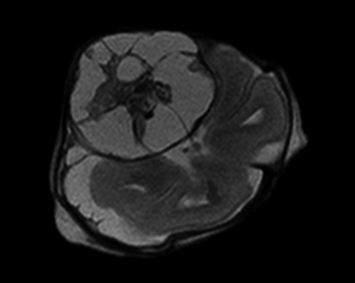
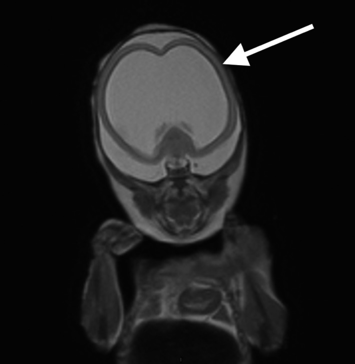
Thirty-four fetuses were evaluated. Sixteen cases were associated with the central nervous system (CNS), five were musculoskeletal, one cardiovascular, one was associated with the urinary tract, and 11 cases had miscellaneous anomalies such as chromosomal aberrations, infections and syndromes. In the 16 cases related to the CNS, both radiologists reported all or some, including the most clinically significant anomalies in 15 (94%; CI 70%-100%) cases. In the 18 non-CNS cases, both radiologists reported all or some, including the most clinically significant anomalies in six (33%; CI 5%-85%) cases. In 21 cases (62%; CI 44%-78%), both radiologists held opinions that were consistent with the autopsy reports. The degree of agreement between the radiologists was high, with a Cohen's Kappa of 0.87.
Postmortem fetal MRI can replace autopsy for second trimester fetuses with CNS anomalies. For non-CNS anomalies, the concordance is lower but postmortem MRI can still be of value when autopsy is not an option.
我们的目的是研究与因胎儿畸形而终止妊娠的胎儿中孕期尸检相比,胎儿磁共振成像(MRI)的准确性。次要目的是比较两位资深放射科医生的 MRI 评估结果。
这是一项前瞻性研究,纳入了 34 例因胎儿畸形而在中孕期终止妊娠的胎儿。所有女性均接受了胎儿死后 MRI 和胎儿尸检。两位资深放射科医生对 MRI 图像进行了独立评估。一位资深病理学家进行了胎儿尸检。MRI 评估与尸检报告的一致性程度以及放射科医生之间的共识程度均进行了评估。
共评估了 34 例胎儿。16 例与中枢神经系统(CNS)有关,5 例与肌肉骨骼系统有关,1 例与心血管系统有关,1 例与泌尿系统有关,11 例有多种异常,如染色体异常、感染和综合征。在与 CNS 相关的 16 例病例中,两位放射科医生均报告了所有或部分病例,包括 15 例(94%;CI 70%-100%)最具临床意义的异常。在 18 例非 CNS 病例中,两位放射科医生均报告了所有或部分病例,包括 6 例(33%;CI 5%-85%)最具临床意义的异常。在 21 例(62%;CI 44%-78%)病例中,两位放射科医生的意见与尸检报告一致。两位放射科医生的意见一致性较高,Cohen's Kappa 值为 0.87。
对于中枢神经系统畸形的中孕期胎儿,死后胎儿 MRI 可替代尸检。对于非 CNS 异常,一致性较低,但当无法进行尸检时,死后 MRI 仍然具有价值。