Department of Paediatric Otolaryngology, Starship Children's Hospital, Auckland, New Zealand.
Department of Anatomy and Medical Imaging, Faculty of Medical and Health Sciences, University of Auckland, Auckland, New Zealand.
Clin Anat. 2019 Sep;32(6):749-761. doi: 10.1002/ca.23343. Epub 2019 Feb 19.
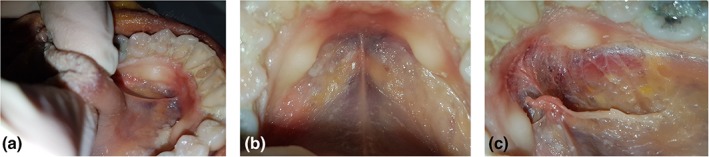
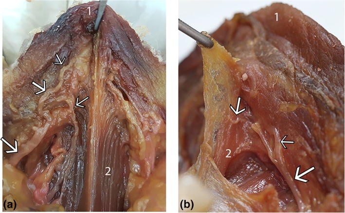
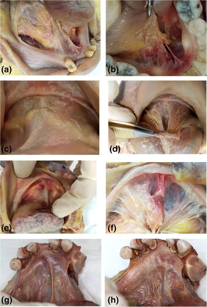
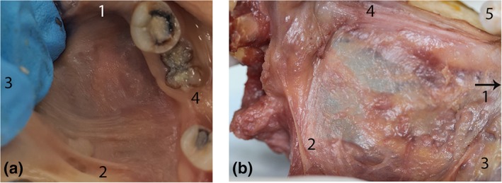
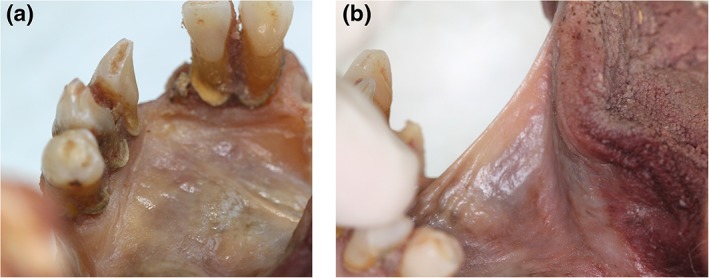
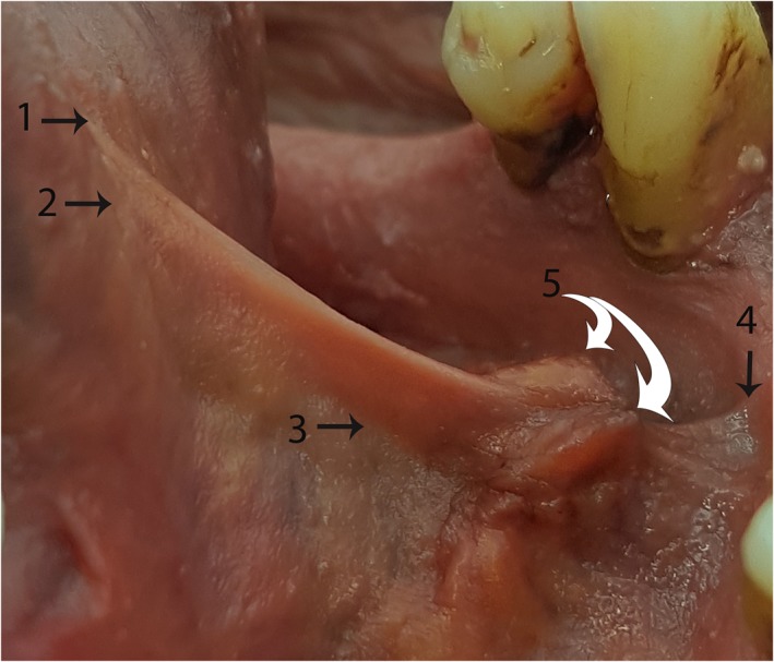
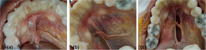
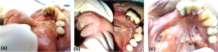
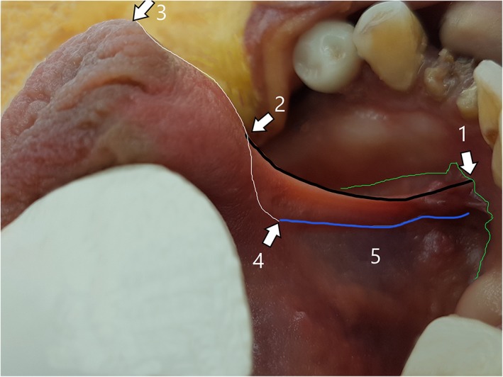
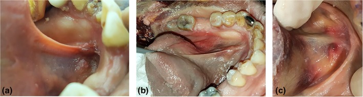
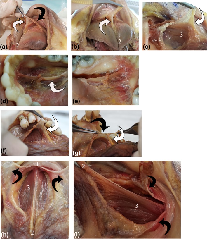
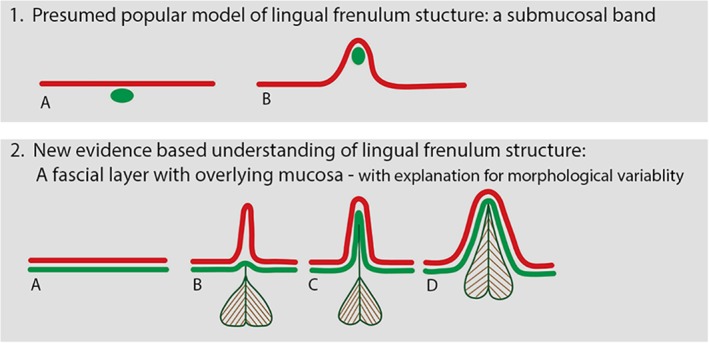
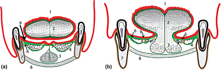
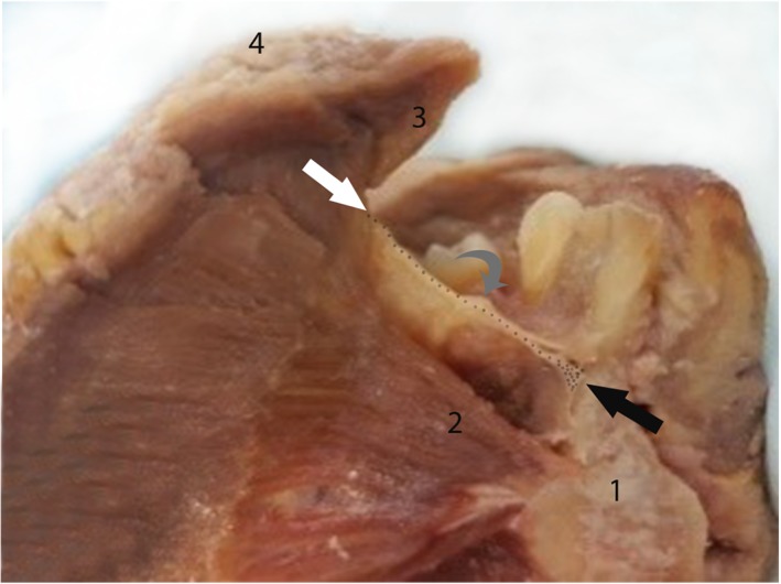
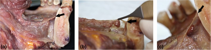
Surgical release of the lingual frenulum (frenotomy) has become an increasingly common procedure, performed from birth through to adulthood. Surprisingly, detailed anatomy of the in-situ lingual frenulum has never been described, and no anatomical basis has been proposed for the individual variability in frenulum morphology. The lingual frenulum is frequently referred to as a "cord" or "submucosal band" of connective tissue, yet there is no evidence to support this anatomical construct. This paper aims to describe the anatomy of the in-situ lingual frenulum and its relationship to floor of mouth structures. Fresh tissue microdissection of the lingual frenulum and floor of mouth was performed on nine adult cadavers with photo-documentation and description of findings. The lingual frenulum is a dynamic structure, formed by a midline fold in a layer of fascia that inserts around the inner arc of the mandible, forming a diaphragm-like structure across the floor of mouth. This fascia is located immediately beneath the oral mucosa, fusing centrally with the connective tissue on the tongue's ventral surface. The sublingual glands and submandibular ducts are enveloped by the fascial layer and anterior genioglossus fibers are suspended beneath it. Lingual nerve branches are located superficially on the ventral surface of the tongue, immediately deep to the fascia. The lingual frenulum is not a discrete midline structure. It is formed by dynamic elevation of a midline fold in the floor of mouth fascia. With this study, the clinical concept of ankyloglossia and its surgical management warrant revision. Clin. Anat. 32:749-761, 2019. © 2019 The Authors. Clinical Anatomy published by Wiley Periodicals, Inc. on behalf of American Association of Clinical Anatomists.
舌系带松解术(系带切开术)已成为一种越来越常见的手术,从出生到成年都在进行。令人惊讶的是,舌系带的原位解剖结构从未被描述过,也没有提出关于系带形态个体差异的解剖学基础。舌系带通常被称为结缔组织的“索”或“黏膜下带”,但没有证据支持这种解剖结构。本文旨在描述原位舌系带及其与口底结构的关系。在九具成人尸体上进行了舌系带和口底的新鲜组织显微解剖,并对发现的结构进行了拍照和描述。舌系带是一种动态结构,由一层筋膜中的中线折叠形成,该筋膜插入下颌骨内弧周围,形成横跨口底的膈状结构。该筋膜位于口腔黏膜下方,在舌腹面的结缔组织中央融合。舌下腺和下颌下腺导管被筋膜层包裹,颏舌肌纤维悬于其下。舌神经分支位于舌腹面的浅层,紧邻筋膜下方。舌系带不是一个独立的中线结构。它是由口底筋膜中线折叠的动态抬高形成的。通过这项研究,临床术语“舌系带过紧”及其手术治疗需要修正。临床解剖学 32:749-761, 2019. © 2019 作者。临床解剖学由 Wiley Periodicals, Inc. 代表美国临床解剖学家协会出版。