• 文献检索
  • 文档翻译
  • 深度研究
  • 学术资讯
  • Suppr Zotero 插件Zotero 插件
  • 邀请有礼
  • 套餐&价格
  • 历史记录
应用&插件
Suppr Zotero 插件Zotero 插件浏览器插件Mac 客户端Windows 客户端微信小程序
定价
高级版会员购买积分包购买API积分包
服务
文献检索文档翻译深度研究API 文档MCP 服务
关于我们
关于 Suppr公司介绍联系我们用户协议隐私条款
关注我们

Suppr 超能文献

核心技术专利:CN118964589B侵权必究
粤ICP备2023148730 号-1Suppr @ 2026

文献检索

告别复杂PubMed语法,用中文像聊天一样搜索,搜遍4000万医学文献。AI智能推荐,让科研检索更轻松。

立即免费搜索

文件翻译

保留排版,准确专业,支持PDF/Word/PPT等文件格式,支持 12+语言互译。

免费翻译文档

深度研究

AI帮你快速写综述,25分钟生成高质量综述,智能提取关键信息,辅助科研写作。

立即免费体验

组织转谷氨酰胺酶介导的 AT1 受体敏化是炎症细胞因子 LIGHT 诱导的高血压的基础。

Tissue Transglutaminase-Mediated AT1 Receptor Sensitization Underlies Pro-inflammatory Cytokine LIGHT-Induced Hypertension.

机构信息

Department of Biochemistry and Molecular Biology, McGovern Medical School at Houston, University of Texas, Houston, Texas, USA.

Department of Nephrology, The First Xiangya Hospital of Central South University, Changsha, Hunan, PRC.

出版信息

Am J Hypertens. 2019 Apr 22;32(5):476-485. doi: 10.1093/ajh/hpz018.

DOI:10.1093/ajh/hpz018
PMID:30715101
原文链接:https://pmc.ncbi.nlm.nih.gov/articles/PMC6475879/
Abstract

BACKGROUND

Although numerous recent studies have shown a strong link between inflammation and hypertension, the underlying mechanisms by which inflammatory cytokines induce hypertension remain to be fully elucidated. Hypertensive disorders are also associated with elevated pressor sensitivity. Tissue transglutaminase (TG2), a potent cross-linking enzyme, is known to be transcriptionally activated by inflammatory cytokines and stabilize angiotensin II (Ang II) receptor AT1 (AT1R) via ubiquitination-preventing posttranslational modification. Here we sought to investigate the TG2-mediated AT1R stabilization in inflammation-induced hypertension and its functional consequences with a focus on receptor abundance and Ang II responsiveness.

METHODS AND RESULTS

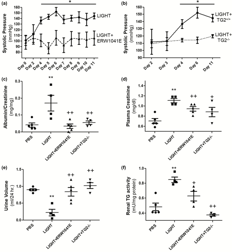
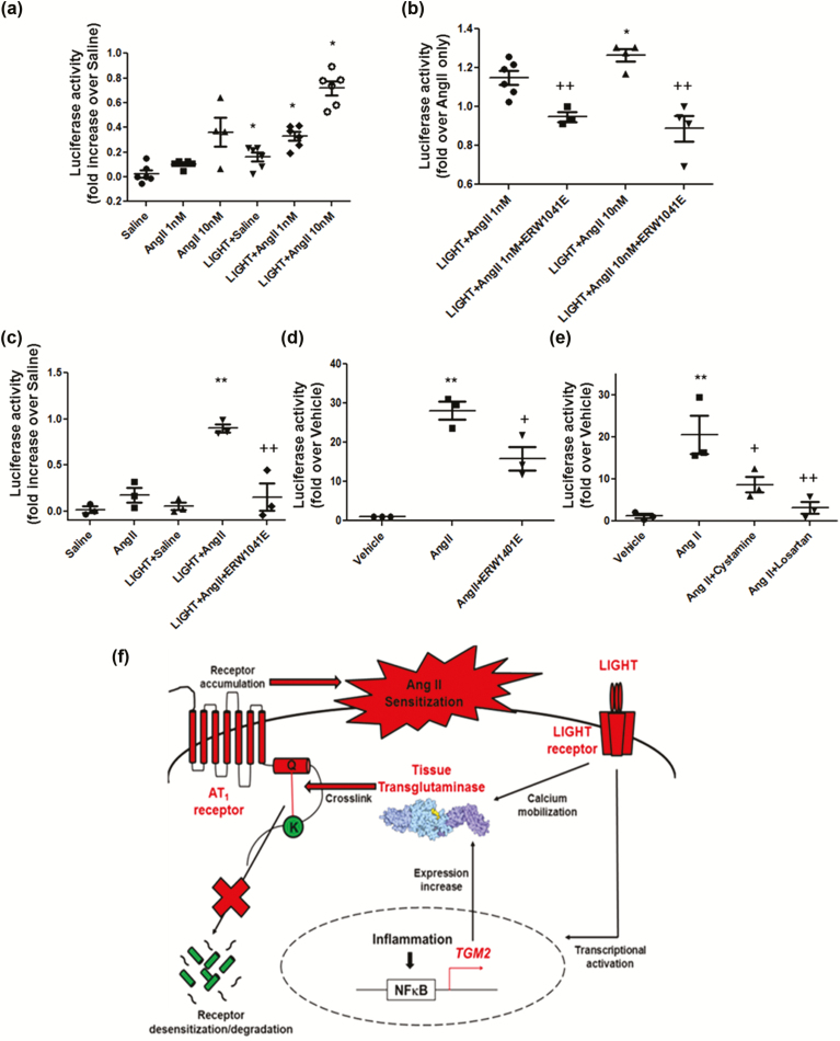
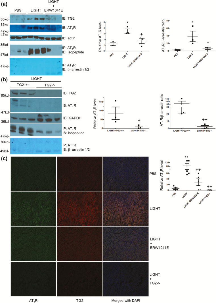
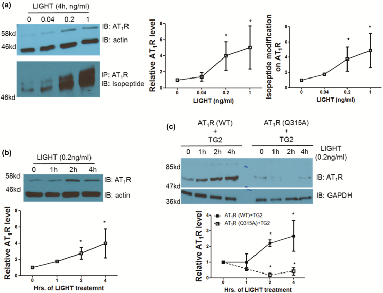
Using an experimental model of inflammation-induced hypertension established by introducing the pro-inflammatory tumor necrosis factor cytokine LIGHT, we provide pharmacologic and genetic evidence that TG2 is required for LIGHT-induced hypertension (systolic pressure on day 6: LIGHT = 152.3 ± 7.4 vs. LIGHT+ERW1041E [TG2 inhibitor] = 105.8 ± 13.1 or LIGHT+TG2-/- = 114.3 ± 4.3 mm Hg, P < 0.05, n = 4-5) and renal compromise (urine albumin/creatinine: LIGHT = 0.17 ± 0.05 vs. LIGHT+ERW1041E = 0.03 ± 0.01 or LIGHT+TG2-/- = 0.06 ± 0.01 mg/mg; plasma creatinine: LIGHT = 1.11 ± 0.04 vs. LIGHT+ERW1041E = 0.94 ± 0.04 or LIGHT+TG2-/- = 0.88 ± 0.09 mg/dl; urine volume: LIGHT = 0.23 ± 0.1 vs. LIGHT+ERW1041E = 0.84 ± 0.13 or LIGHT+TG2-/- = 1.02 ± 0.09 ml/24 hour on day 14, P < 0.05, n = 4-5). Our mechanistic studies showed that the TG2-mediated AT1R modification and accumulation (relative renal AT1R level: phosphate-buffered saline [PBS] = 1.23 ± 0.22, LIGHT = 3.49 ± 0.37, and LIGHT+ERW1041E = 1.77 ± 0.46, P < 0.05, n = 3; LIGHT+TG2+/+ = 85.28 ± 36.11 vs. LIGHT+TG2-/- = 7.01 ± 5.68, P < 0.05, n = 3) induced by LIGHT is associated with abrogated β-arrestin binding (AT1R/associated β-arrestin ratio: PBS = 2.62 ± 1.07, LIGHT = 38.60 ± 13.91, and LIGHT+ERW1041E = 6.97 ± 2.91, P < 0.05, n = 3; LIGHT+TG2+/+ = 66.43 ± 44.81 vs. LIGHT+TG2-/- = 2.45 ± 1.78, P < 0.01, n = 3) and could be found in renal medulla tubules of kidneys (relative tubular AT1R level: PBS = 5.91 ± 2.93, LIGHT = 92.82 ± 19.54, LIGHT+ERW1041E = 28.49 ± 11.65, and LIGHT+TG2-/- = 0.14 ± 0.10, P < 0.01, n = 5) and the blood vasculature (relative vascular AT1R level: PBS = 0.70 ± 0.30, LIGHT = 13.75 ± 2.49, and LIGHT+ERW1041E = 3.28 ± 0.87, P < 0.01, n = 3), 2 of the tissues highly related to the genesis of hypertension. Our in vitro cellular assays showed that LIGHT stimulation triggered a rapid TG2-dependent increase in the abundance of AT1Rs (relative AT1R level after 2-hour LIGHT treatment: AT1R (WT)+TG2 = 2.21 ± 0.23, AT1R (Q315A)+TG2 = 0.18 ± 0.23, P < 0.05 vs. starting point = 1, n = 2) and downstream calcium signaling (fold increase in NFAT-driven luciferase activity: Saline = 0.02 ± 0.03, Ang II = 0.17 ± 0.08, LIGHT = 0.05 ± 0.04, LIGHT+Ang II = 0.90 ± 0.04 (P < 0.01 vs. Ang II), and LIGHT+Ang II+ERW1041E = 0.15 ± 0.15 (P < 0.01 vs. LIGHT+Ang II), n = 3).

CONCLUSIONS

Our data indicate an essential and systemic role for TG2 in bridging inflammation to hypertension via its posttranslational modifications stabilizing AT1 receptor and sensitizing Ang II. Our findings also suggest that TG2 inhibitors could be used as a novel group of cardiovascular agents.

摘要

背景

尽管最近有大量研究表明炎症与高血压之间存在很强的关联,但炎症细胞因子引起高血压的确切机制仍未完全阐明。高血压疾病也与血压升高的敏感性有关。组织转谷氨酰胺酶(TG2)是一种有效的交联酶,已知可被炎症细胞因子转录激活,并通过阻止翻译后修饰稳定血管紧张素 II(Ang II)受体 AT1(AT1R)。在这里,我们试图研究 TG2 在炎症诱导的高血压中的作用及其对受体丰度和 Ang II 反应性的影响。

方法和结果

我们使用了一种通过引入促炎肿瘤坏死因子细胞因子 LIGHT 建立的炎症诱导的高血压实验模型,提供了药理学和遗传学证据,表明 TG2 是 LIGHT 诱导的高血压(第 6 天收缩压:LIGHT = 152.3 ± 7.4 与 LIGHT+ERW1041E [TG2 抑制剂] = 105.8 ± 13.1 或 LIGHT+TG2-/- = 114.3 ± 4.3 mmHg,P < 0.05,n = 4-5)和肾脏损伤(尿白蛋白/肌酐:LIGHT = 0.17 ± 0.05 与 LIGHT+ERW1041E = 0.03 ± 0.01 或 LIGHT+TG2-/- = 0.06 ± 0.01 mg/mg;血浆肌酐:LIGHT = 1.11 ± 0.04 与 LIGHT+ERW1041E = 0.94 ± 0.04 或 LIGHT+TG2-/- = 0.88 ± 0.09 mg/dl;尿体积:LIGHT = 0.23 ± 0.1 与 LIGHT+ERW1041E = 0.84 ± 0.13 或 LIGHT+TG2-/- = 1.02 ± 0.09 ml/24 小时,P < 0.05,n = 4-5)所必需的。我们的机制研究表明,TG2 介导的 AT1R 修饰和积累(相对肾 AT1R 水平:磷酸盐缓冲盐水 [PBS] = 1.23 ± 0.22,LIGHT = 3.49 ± 0.37,和 LIGHT+ERW1041E = 1.77 ± 0.46,P < 0.05,n = 3;LIGHT+TG2+/+ = 85.28 ± 36.11 与 LIGHT+TG2-/- = 7.01 ± 5.68,P < 0.05,n = 3)与结合β-抑制蛋白的减少(AT1R/结合β-抑制蛋白比值:PBS = 2.62 ± 1.07,LIGHT = 38.60 ± 13.91,和 LIGHT+ERW1041E = 6.97 ± 2.91,P < 0.05,n = 3;LIGHT+TG2+/+ = 66.43 ± 44.81 与 LIGHT+TG2-/- = 2.45 ± 1.78,P < 0.01,n = 3)有关,并且可以在肾脏髓质肾小管中发现(相对肾小管 AT1R 水平:PBS = 5.91 ± 2.93,LIGHT = 92.82 ± 19.54,LIGHT+ERW1041E = 28.49 ± 11.65,和 LIGHT+TG2-/- = 0.14 ± 0.10,P < 0.01,n = 5)和血管(相对血管 AT1R 水平:PBS = 0.70 ± 0.30,LIGHT = 13.75 ± 2.49,和 LIGHT+ERW1041E = 3.28 ± 0.87,P < 0.01,n = 3),这两个组织与高血压的发生密切相关。我们的体外细胞试验表明,LIGHT 刺激触发了 AT1R 丰度的快速 TG2 依赖性增加(2 小时 LIGHT 处理后的相对 AT1R 水平:AT1R(WT)+TG2 = 2.21 ± 0.23,AT1R(Q315A)+TG2 = 0.18 ± 0.23,P < 0.05 与起始点 = 1,n = 2)和下游钙信号(NFAT 驱动的荧光素酶活性的倍数增加:盐水 = 0.02 ± 0.03,Ang II = 0.17 ± 0.08,LIGHT = 0.05 ± 0.04,LIGHT+Ang II = 0.90 ± 0.04(P < 0.01 与 Ang II),和 LIGHT+Ang II+ERW1041E = 0.15 ± 0.15(P < 0.01 与 LIGHT+Ang II),n = 3)。

结论

我们的数据表明,TG2 在通过其稳定 AT1 受体和敏化 Ang II 的翻译后修饰将炎症与高血压联系起来方面发挥了重要的系统性作用。我们的发现还表明,TG2 抑制剂可作为一类新的心血管药物。

https://cdn.ncbi.nlm.nih.gov/pmc/blobs/5076/6475879/fcb054dcfe9f/hpz018f0004.jpg
https://cdn.ncbi.nlm.nih.gov/pmc/blobs/5076/6475879/58fdafa10fb2/hpz018f0001.jpg
https://cdn.ncbi.nlm.nih.gov/pmc/blobs/5076/6475879/24a33d4a433b/hpz018f0002.jpg
https://cdn.ncbi.nlm.nih.gov/pmc/blobs/5076/6475879/f5c9be1037de/hpz018f0003.jpg
https://cdn.ncbi.nlm.nih.gov/pmc/blobs/5076/6475879/fcb054dcfe9f/hpz018f0004.jpg

相似文献

1
Tissue Transglutaminase-Mediated AT1 Receptor Sensitization Underlies Pro-inflammatory Cytokine LIGHT-Induced Hypertension.组织转谷氨酰胺酶介导的 AT1 受体敏化是炎症细胞因子 LIGHT 诱导的高血压的基础。
Am J Hypertens. 2019 Apr 22;32(5):476-485. doi: 10.1093/ajh/hpz018.
2
Inflammation, Autoimmunity, and Hypertension: The Essential Role of Tissue Transglutaminase.炎症、自身免疫和高血压:组织转谷氨酰胺酶的重要作用。
Am J Hypertens. 2017 Aug 1;30(8):756-764. doi: 10.1093/ajh/hpx027.
3
Tissue transglutaminase contributes to the pathogenesis of preeclampsia and stabilizes placental angiotensin receptor type 1 by ubiquitination-preventing isopeptide modification.组织转谷氨酰胺酶通过泛素化防止异肽修饰稳定胎盘血管紧张素受体 1,从而促进子痫前期的发病机制。
Hypertension. 2014 Feb;63(2):353-61. doi: 10.1161/HYPERTENSIONAHA.113.02361. Epub 2013 Nov 4.
4
Transglutaminase is a Critical Link Between Inflammation and Hypertension.转谷氨酰胺酶是炎症与高血压之间的关键纽带。
J Am Heart Assoc. 2016 Jun 30;5(7):e003730. doi: 10.1161/JAHA.116.003730.
5
Vasopressor meets vasodepressor: The AT1-B2 receptor heterodimer.血管加压素遇见血管舒张素:AT1-B2 受体异二聚体。
Biochem Pharmacol. 2014 Apr 1;88(3):284-90. doi: 10.1016/j.bcp.2014.01.019. Epub 2014 Jan 22.
6
Purinergic P2Y6 receptors heterodimerize with angiotensin AT1 receptors to promote angiotensin II-induced hypertension.嘌呤能P2Y6受体与血管紧张素AT1受体形成异源二聚体,以促进血管紧张素II诱导的高血压。
Sci Signal. 2016 Jan 19;9(411):ra7. doi: 10.1126/scisignal.aac9187.
7
Central Renin-Angiotensin System Activation and Inflammation Induced by High-Fat Diet Sensitize Angiotensin II-Elicited Hypertension.高脂饮食诱导的中枢肾素-血管紧张素系统激活和炎症会使血管紧张素II引发的高血压更易发生。
Hypertension. 2016 Jan;67(1):163-70. doi: 10.1161/HYPERTENSIONAHA.115.06263. Epub 2015 Nov 16.
8
Angiotensin II mediates angiotensin converting enzyme type 2 internalization and degradation through an angiotensin II type I receptor-dependent mechanism.血管紧张素II通过血管紧张素II 1型受体依赖性机制介导血管紧张素转换酶2的内化和降解。
Hypertension. 2014 Dec;64(6):1368-1375. doi: 10.1161/HYPERTENSIONAHA.114.03743. Epub 2014 Sep 15.
9
Captopril avoids hypertension, the increase in plasma angiotensin II but increases angiotensin 1-7 and angiotensin II-induced perfusion pressure in isolated kidney in SHR.卡托普利可避免高血压、血浆血管紧张素II升高,但会增加自发性高血压大鼠离体肾脏中血管紧张素1-7及血管紧张素II诱导的灌注压。
Auton Autacoid Pharmacol. 2012 Oct;32(3 Pt 4):61-9. doi: 10.1111/aap.12001.
10
Interaction of Angiotensin II AT1 Receptors with Purinergic P2X Receptors in Regulating Renal Afferent Arterioles in Angiotensin II-Dependent Hypertension.血管紧张素 II AT1 受体与嘌呤能 P2X 受体在血管紧张素 II 依赖性高血压调节肾入球小动脉中的相互作用。
Int J Mol Sci. 2023 Jul 13;24(14):11413. doi: 10.3390/ijms241411413.

引用本文的文献

1
Renin-Angiotensin System Autoantibody Network in Parkinson's Disease Patients.帕金森病患者的肾素-血管紧张素系统自身抗体网络
Antioxidants (Basel). 2025 Jun 10;14(6):706. doi: 10.3390/antiox14060706.
2
Serum angiotensin type-1 receptor autoantibodies and neurofilament light chain as markers of neuroaxonal damage in post-COVID patients.血清1型血管紧张素受体自身抗体和神经丝轻链作为新冠康复患者神经轴突损伤的标志物
Front Immunol. 2025 Apr 22;16:1571027. doi: 10.3389/fimmu.2025.1571027. eCollection 2025.
3
Non-HLA angiotensin-type-1 receptor autoantibodies mediate the long-term loss of grafted neurons in Parkinson's disease models.

本文引用的文献

1
Pharmacologic inhibition of the enzymatic effects of tissue transglutaminase reduces cardiac fibrosis and attenuates cardiomyocyte hypertrophy following pressure overload.药物抑制组织转谷氨酰胺酶的酶作用可减少压力超负荷后的心肌纤维化并减轻心肌细胞肥大。
J Mol Cell Cardiol. 2018 Apr;117:36-48. doi: 10.1016/j.yjmcc.2018.02.016. Epub 2018 Mar 2.
2
Inflammation, Autoimmunity, and Hypertension: The Essential Role of Tissue Transglutaminase.炎症、自身免疫和高血压:组织转谷氨酰胺酶的重要作用。
Am J Hypertens. 2017 Aug 1;30(8):756-764. doi: 10.1093/ajh/hpx027.
3
Transglutaminase is a Critical Link Between Inflammation and Hypertension.
非 HLA 血管紧张素受体 1 自身抗体介导帕金森病模型中移植神经元的长期丢失。
Stem Cell Res Ther. 2024 May 12;15(1):138. doi: 10.1186/s13287-024-03751-y.
4
S-Nitrosylation of Tissue Transglutaminase in Modulating Glycolysis, Oxidative Stress, and Inflammatory Responses in Normal and Indoxyl-Sulfate-Induced Endothelial Cells.组织转谷氨酰胺酶的 S-亚硝基化在调节正常和硫酸吲哚酚诱导的内皮细胞糖酵解、氧化应激和炎症反应中的作用。
Int J Mol Sci. 2023 Jun 30;24(13):10935. doi: 10.3390/ijms241310935.
5
Angiotensin type-1 receptor and ACE2 autoantibodies in Parkinson´s disease.帕金森病中的血管紧张素1型受体和血管紧张素转换酶2自身抗体。
NPJ Parkinsons Dis. 2022 Jun 14;8(1):76. doi: 10.1038/s41531-022-00340-9.
6
Autoantibodies against ACE2 and angiotensin type-1 receptors increase severity of COVID-19.抗血管紧张素转换酶 2(ACE2)和血管紧张素 I 型受体自身抗体增加 COVID-19 严重程度。
J Autoimmun. 2021 Aug;122:102683. doi: 10.1016/j.jaut.2021.102683. Epub 2021 Jun 11.
7
Increased Transglutaminase 2 Expression and Activity in Rodent Models of Obesity/Metabolic Syndrome and Aging.肥胖/代谢综合征和衰老啮齿动物模型中转谷氨酰胺酶2表达及活性增加
Front Physiol. 2020 Sep 15;11:560019. doi: 10.3389/fphys.2020.560019. eCollection 2020.
8
NADPH Oxidase Inhibition in Fibrotic Pathologies.NADPH 氧化酶在纤维化病理中的抑制作用。
Antioxid Redox Signal. 2020 Aug 20;33(6):455-479. doi: 10.1089/ars.2020.8032. Epub 2020 Mar 4.
9
Age-Dependent Impairment in Endothelial Function and Arterial Stiffness in Former High Class Male Athletes Is No Different to That in Men With No History of Physical Training.年龄相关性内皮功能障碍和动脉僵硬度在曾为高水平男性运动员和无体力训练史男性中的差异无统计学意义。
J Am Heart Assoc. 2019 Sep 17;8(18):e012670. doi: 10.1161/JAHA.119.012670. Epub 2019 Sep 12.
转谷氨酰胺酶是炎症与高血压之间的关键纽带。
J Am Heart Assoc. 2016 Jun 30;5(7):e003730. doi: 10.1161/JAHA.116.003730.
4
Endostatin and transglutaminase 2 are involved in fibrosis of the aging kidney.内皮抑素和转谷氨酰胺酶2与衰老肾脏的纤维化有关。
Kidney Int. 2016 Jun;89(6):1281-92. doi: 10.1016/j.kint.2016.01.030. Epub 2016 Apr 14.
5
Role of Inflammation and Immunity in Hypertension: Recent Epidemiological, Laboratory, and Clinical Evidence.炎症与免疫在高血压中的作用:近期的流行病学、实验室及临床证据
Curr Hypertens Rep. 2016 Mar;18(3):21. doi: 10.1007/s11906-016-0628-7.
6
Central Renin-Angiotensin System Activation and Inflammation Induced by High-Fat Diet Sensitize Angiotensin II-Elicited Hypertension.高脂饮食诱导的中枢肾素-血管紧张素系统激活和炎症会使血管紧张素II引发的高血压更易发生。
Hypertension. 2016 Jan;67(1):163-70. doi: 10.1161/HYPERTENSIONAHA.115.06263. Epub 2015 Nov 16.
7
Brief serotonin exposure initiates arteriolar inward remodeling processes in vivo that involve transglutaminase activation and actin cytoskeleton reorganization.短暂的血清素暴露在体内引发小动脉内向重塑过程,这涉及转谷氨酰胺酶激活和肌动蛋白细胞骨架重组。
Am J Physiol Heart Circ Physiol. 2016 Jan 15;310(2):H188-98. doi: 10.1152/ajpheart.00666.2015. Epub 2015 Nov 13.
8
Elevated Serum Level of Interleukin 17 in a Population With Prehypertension.高血压前期人群中白细胞介素17血清水平升高
J Clin Hypertens (Greenwich). 2015 Oct;17(10):770-4. doi: 10.1111/jch.12612. Epub 2015 Jul 3.
9
Inflammation, immunity, and hypertensive end-organ damage.炎症、免疫与高血压性靶器官损害。
Circ Res. 2015 Mar 13;116(6):1022-33. doi: 10.1161/CIRCRESAHA.116.303697.
10
Transglutaminase regulation of cell function.转谷氨酰胺酶对细胞功能的调节。
Physiol Rev. 2014 Apr;94(2):383-417. doi: 10.1152/physrev.00019.2013.