Department of Medicine, University of Cambridge, MRC Laboratory of Molecular Biology, UK.
MRC Laboratory of Molecular Biology, Cambridge, UK.
FEBS J. 2019 Apr;286(8):1543-1560. doi: 10.1111/febs.14772. Epub 2019 Feb 16.
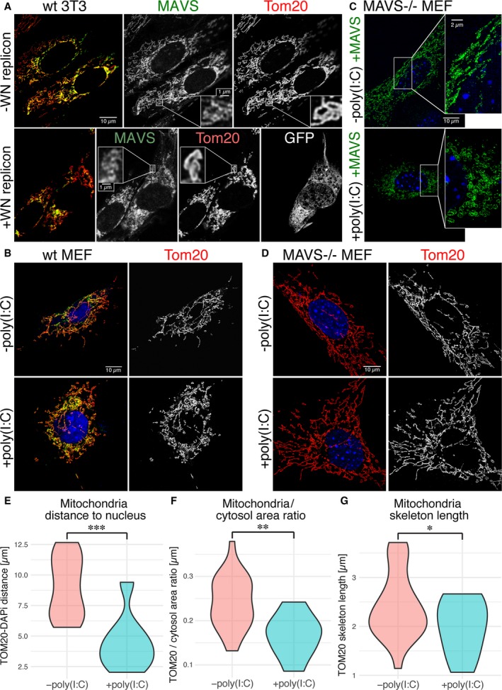
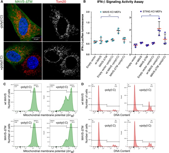
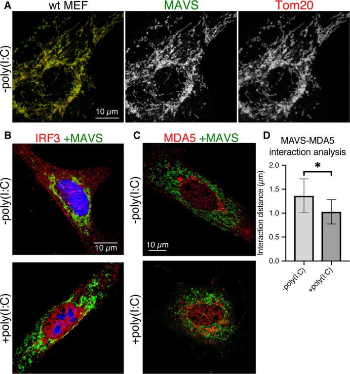
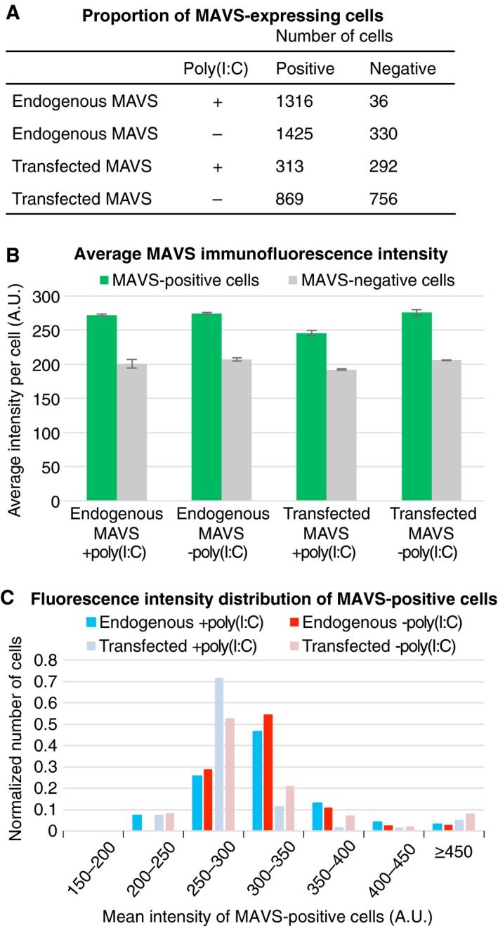
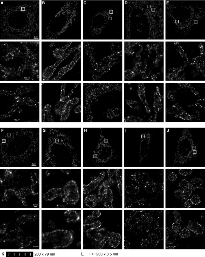
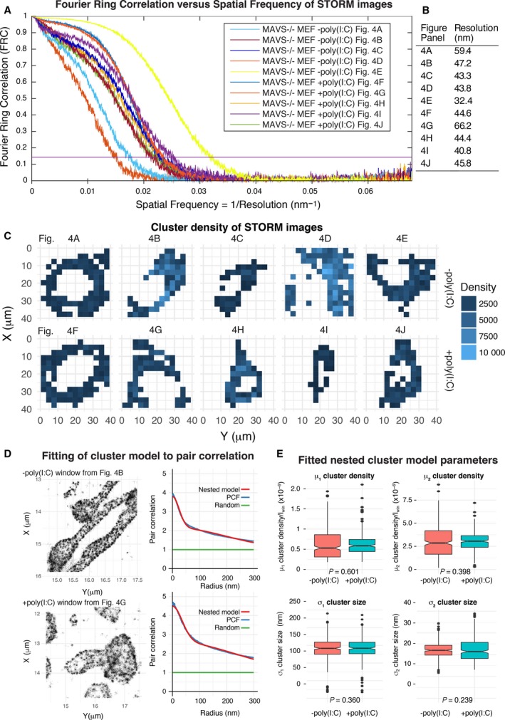
Double-stranded RNA (dsRNA) is a potent proinflammatory signature of viral infection and is sensed primarily by RIG-I-like receptors (RLRs). Oligomerization of RLRs following binding to cytosolic dsRNA activates and nucleates self-assembly of the mitochondrial antiviral-signaling protein (MAVS). In the current signaling model, the caspase recruitment domains of MAVS form helical fibrils that self-propagate like prions to promote signaling complex assembly. However, there is no conclusive evidence that MAVS forms fibrils in cells or with the transmembrane anchor present. We show here with super-resolution light microscopy that MAVS activation by dsRNA induces mitochondrial membrane remodeling. Quantitative image analysis at imaging resolutions as high as 32 nm shows that in the cellular context, MAVS signaling complexes and the fibrils within them are smaller than 80 nm. The transmembrane domain of MAVS is required for its membrane remodeling, interferon signaling, and proapoptotic activities. We conclude that membrane tethering of MAVS restrains its polymerization and contributes to mitochondrial remodeling and apoptosis upon dsRNA sensing.
双链 RNA(dsRNA)是病毒感染的一种有效的促炎特征,主要由 RIG-I 样受体(RLR)感知。RLR 结合细胞质 dsRNA 后寡聚化,激活并引发抗病毒信号蛋白(MAVS)的线粒体自我组装。在当前的信号模型中,MAVS 的半胱天冬酶募集结构域形成螺旋状纤维,像朊病毒一样自我传播,以促进信号复合物的组装。然而,尚无确凿证据表明 MAVS 在细胞中或与跨膜锚存在的情况下形成纤维。我们在这里通过超分辨率光显微镜显示,dsRNA 激活 MAVS 诱导线粒体膜重塑。在高达 32nm 的成像分辨率下进行定量图像分析表明,在细胞环境中,MAVS 信号复合物及其内部的纤维小于 80nm。MAVS 的跨膜结构域是其膜重塑、干扰素信号和促凋亡活性所必需的。我们得出结论,MAVS 的膜束缚抑制了其聚合,并有助于 dsRNA 感应时的线粒体重塑和凋亡。