Aljarbou Fahd A, Aldosimani Mazen, Althumairy Riyadh I, Alhezam Abdullah A, Aldawsari Abdullah I
Department of Restorative Dental Sciences, Division of Endodontics, College of Dentistry, King Saud University, Riyadh, Kingdom of Saudi Arabia. E-mail.
Saudi Med J. 2019 Feb;40(2):189-194. doi: 10.15537/smj.2019.2.23602.
To evaluate the relationship of the first and second mandibular molar roots to the inferior alveolar canal (IAC) and cortical plates using cone beam computed tomography (CBCT) in the Saudi population. Methods: Scans of 60 patients were collected retrospectively from the dental hospital database in King Saud University, Riyadh, Kingdom of Saudi Arabia. Measurements of the right and left first and second mandibular molars for each dental root and the mandibular bone thickness were determined. The position of the IAC was estimated using axial, coronal, and sagittal views. Three examiners performed the measurements independently.
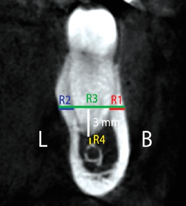
The mean distance between the root apices of the mandibular molars and the IAC ranged from 1.68-4.79 mm, whereas the mean distance from the outer surface of the buccal cortical plate to the buccal root surface ranged from 2.33-6.72 mm. Similarly, the mean distance from the outer surface of the lingual cortical plate to the lingual root surface ranged from 2.62-4.80 mm. Finally, the mean distance from the outer surface of the lingual cortical plate to the outer surface of the buccal cortical plate was 11.93-13.19 mm. Conclusion: The measurements reported in this study may be of value to practitioners treating Saudi patients, as they need to be familiar with the distance of the mandibular first and second molars in relation to the IAC and surrounding cortical plates to accurately assess and plan endodontic surgeries, surgical extractions, and implant placements.
利用锥形束计算机断层扫描(CBCT)评估沙特人群中下颌第一和第二磨牙牙根与下牙槽管(IAC)及皮质骨板的关系。方法:回顾性收集沙特阿拉伯利雅得国王沙特大学牙科医院数据库中60例患者的扫描数据。测定每颗牙左右下颌第一和第二磨牙各牙根以及下颌骨厚度。利用轴向、冠状和矢状位视图估计下牙槽管的位置。由三名检查者独立进行测量。结果:下颌磨牙根尖与下牙槽管的平均距离为1.68 - 4.79毫米,而颊侧皮质骨板外表面至颊侧牙根表面的平均距离为2.33 - 6.72毫米。同样,舌侧皮质骨板外表面至舌侧牙根表面的平均距离为2.62 - 4.80毫米。最后,舌侧皮质骨板外表面至颊侧皮质骨板外表面的平均距离为11.93 - 13.19毫米。结论:本研究报告的测量结果可能对治疗沙特患者的从业者有价值,因为他们需要熟悉下颌第一和第二磨牙相对于下牙槽管及周围皮质骨板的距离,以便准确评估和规划牙髓手术、外科拔牙及种植体植入。