School of Rehabilitation Science, Shanghai University of Traditional Chinese Medicine, Shanghai, China.
Key Laboratory of Hand Reconstruction, Ministry of Health, Shanghai, China.
Neural Plast. 2019 Jan 8;2019:7381609. doi: 10.1155/2019/7381609. eCollection 2019.
Neuropathic pain after brachial plexus injury remains an increasingly prevalent and intractable disease due to inadequacy of satisfactory treatment strategies. A detailed mapping of cortical regions concerning the brain plasticity was the first step of therapeutic intervention. However, the specific mapping research of brachial plexus pain was limited. We aimed to provide some localization information about the brain plasticity changes after brachial plexus pain in this preliminary study.
24 Sprague-Dawley rats received complete brachial plexus avulsion with neuropathic pain on the right forelimb successfully. Through functional imaging of both resting-state and block-design studies, we compared the amplitude of low-frequency fluctuations (ALFF) of premodeling and postmodeling groups and the changes of brain activation when applying sensory stimulation.
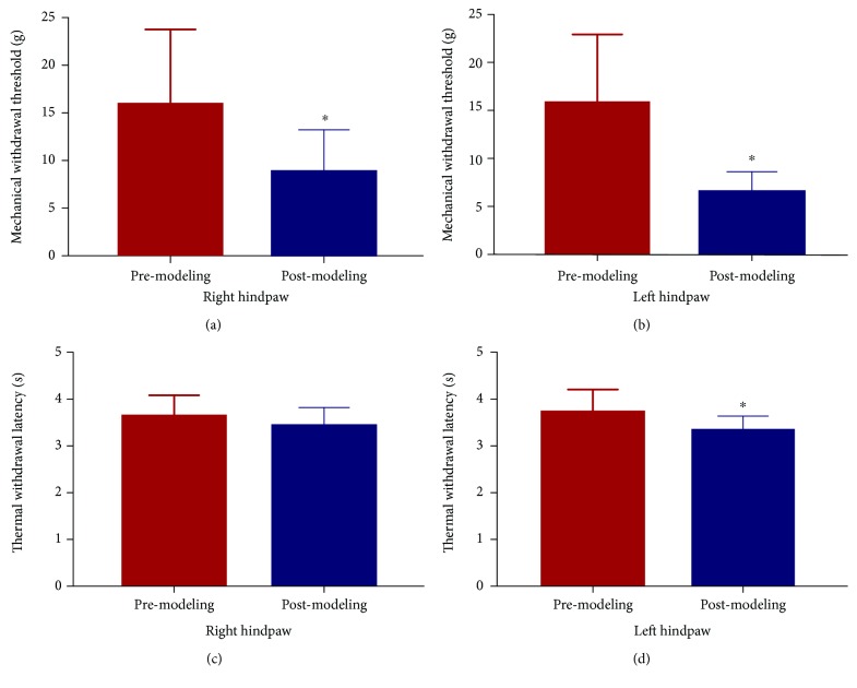
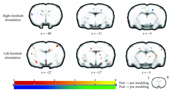
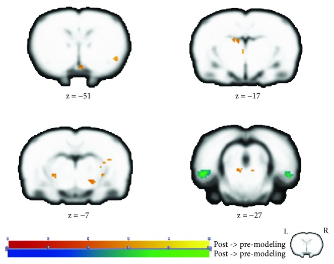
The postmodeling group showed significant decreases on the mechanical withdrawal threshold (MWT) in the bilateral hindpaws and thermal withdrawal latency (TWL) in the left hindpaw than the premodeling group ( < 0.05). The amplitude of low-frequency fluctuations (ALFF) of the postmodeling group manifested increases in regions of the left anterodorsal hippocampus, left mesencephalic region, left dorsal midline thalamus, and so on. Decreased ALFF was observed in the bilateral entorhinal cortex compared to that of the premodeling group. The results of block-design scan showed significant differences in regions including the limbic/paralimbic system and somatosensory cortex.
We concluded that the entorhinal-hippocampus pathway, which was part of the Papez circuit, was involved in the functional integrated areas of brachial plexus pain processing. The regions in the "pain matrix" showed expected activation when applying instant nociceptive stimulus but remained silent in the resting status. This research confirmed the involvement of cognitive function, which brought novel information to the potential new therapy for brachial plexus pain.
臂丛神经损伤后引起的神经性疼痛是一种日益普遍且难以治疗的疾病,这主要是由于缺乏令人满意的治疗策略。详细绘制与大脑可塑性相关的皮质区域图谱是治疗干预的第一步。然而,针对臂丛神经痛的特定映射研究是有限的。在这项初步研究中,我们旨在提供一些关于臂丛神经痛后大脑可塑性变化的定位信息。
24 只 Sprague-Dawley 大鼠成功构建右侧前肢臂丛神经完全撕脱伴有神经性疼痛的动物模型。通过静息态和阻断设计研究的功能成像,我们比较了建模前组和建模后组的低频振幅(ALFF),以及在施加感觉刺激时大脑激活的变化。
建模后组双侧后肢机械缩足反射阈值(MWT)和左侧后肢热缩足潜伏期(TWL)均显著低于建模前组(<0.05)。建模后组的低频振幅(ALFF)表现为左侧前背海马、左侧中脑、左侧背中线丘脑等区域的增加。与建模前组相比,双侧内嗅皮层的 ALFF 降低。阻断设计扫描的结果显示,包括边缘/旁边缘系统和躯体感觉皮层在内的区域存在显著差异。
我们得出结论,内嗅-海马通路,作为 Papez 回路的一部分,参与了臂丛神经痛处理的功能整合区域。“疼痛矩阵”中的区域在施加即时伤害性刺激时表现出预期的激活,但在静息状态下保持沉默。这项研究证实了认知功能的参与,为臂丛神经痛的潜在新疗法提供了新的信息。