Department of Ophthalmology, Osaka University Graduate School of Medicine, Suita, Japan.
Sci Rep. 2019 Feb 7;9(1):1561. doi: 10.1038/s41598-018-38248-1.
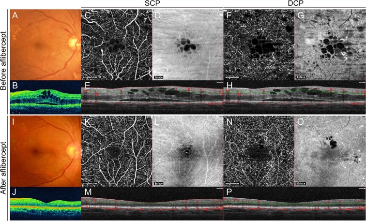
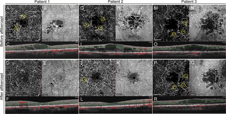
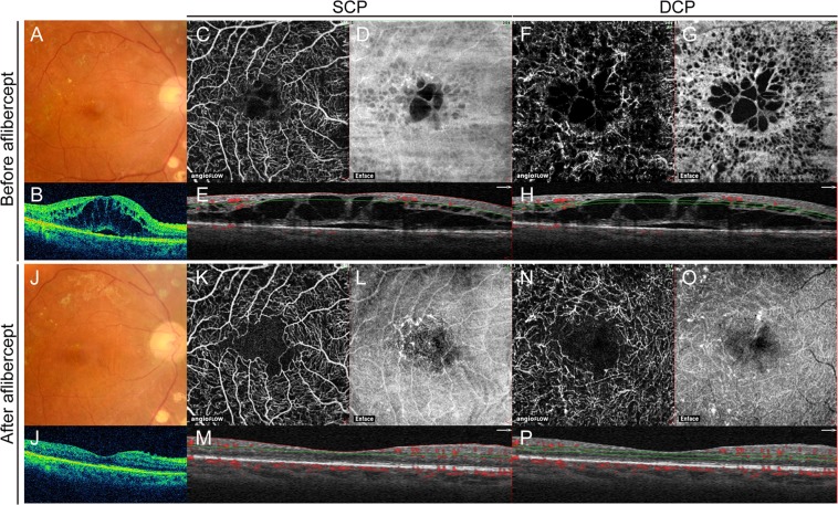
We investigated changes in retinal vascular area and the foveal avascular zone (FAZ) after intravitreal aflibercept in diabetic macular edema (DME) and the association of these changes with visual outcomes. The retinal vascular area in the superficial capillary plexus (SCP) and the deep capillary plexus (DCP) and the FAZ area were measured using optical coherence tomography angiography (OCTA) in 23 eyes of 23 patients with DME, before and after intravitreal aflibercept. Overall, there was no significant change in retinal vascular area or FAZ. Better BCVA after treatment was significantly associated with larger retinal vascular area in the SCP and the DCP, both at baseline (R = 0.512, P < 0.001 and R = 0.361, P = 0.002, respectively) and after intravitreal aflibercept (R = 0.717, P < 0.001 and R = 0.618, P < 0.001, respectively). MAs were observed in the DCP in 20 eyes (87%), but only detected in four eyes (17%) in the SCP before treatment. The number of eyes with MAs in the DCP significantly decreased to 13 (57%) after treatment (P = 0.049). The persistence of DME was associated with persistent MAs (P = 0.019) and less visual gain (P = 0.031) following treatment. Thus, preserving retinal perfusion and the resolution of MAs are associated with better vision and resolution of the DME after intravitreal aflibercept.
我们研究了玻璃体内注射阿柏西普后糖尿病性黄斑水肿(DME)患者视网膜血管面积和中心凹无血管区(FAZ)的变化,以及这些变化与视力结果的关系。使用光学相干断层扫描血管造影(OCTA)测量了 23 例 DME 患者 23 只眼治疗前后浅层毛细血管丛(SCP)和深层毛细血管丛(DCP)的视网膜血管面积和 FAZ 面积。总的来说,视网膜血管面积或 FAZ 没有显著变化。治疗后最佳矫正视力(BCVA)的提高与 SCP 和 DCP 中视网膜血管面积的增大显著相关,基线时(R=0.512,P<0.001 和 R=0.361,P=0.002)以及玻璃体内注射阿柏西普后(R=0.717,P<0.001 和 R=0.618,P<0.001)。在 20 只眼(87%)的 DCP 中观察到微动脉瘤(MA),但在治疗前的 SCP 中仅在 4 只眼(17%)中检测到。治疗后 DCP 中存在 MA 的眼数显著减少至 13 只(57%)(P=0.049)。DME 的持续存在与治疗后持续存在 MA(P=0.019)和视力增益较少(P=0.031)相关。因此,玻璃体内注射阿柏西普后保留视网膜灌注和 MA 的消退与更好的视力和 DME 的消退相关。