Wellcome Centre for Integrative Neuroimaging, FMRIB, Department of Clinical Neurosciences, University of Oxford, Oxford, UK.
Institute for Biomedical Engineering, ETH Zurich and University of Zurich, Zurich, Switzerland.
Magn Reson Med. 2019 Jun;81(6):3745-3753. doi: 10.1002/mrm.27664. Epub 2019 Feb 8.
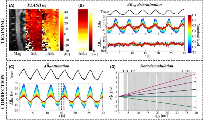
Spinal cord MRI at ultrahigh field is hampered by time-varying magnetic fields associated with the breathing cycle, giving rise to ghosting artifacts in multi-shot acquisitions. Here, we suggest a correction approach based on linking the signal from a respiratory bellows to field changes inside the spinal cord. The information is used to correct the data at the image reconstruction level.
The correction was demonstrated in the context of multi-shot T2*-weighted imaging of the cervical spinal cord at 7T. A respiratory trace was acquired during a high-resolution multi-echo gradient-echo sequence, used for structural imaging and quantitative T2* mapping, and a multi-shot EPI time series, as would be suitable for fMRI. The coupling between the trace and the breathing-induced fields was determined by a short calibration scan in each individual. Images were reconstructed with and without trace-based correction.
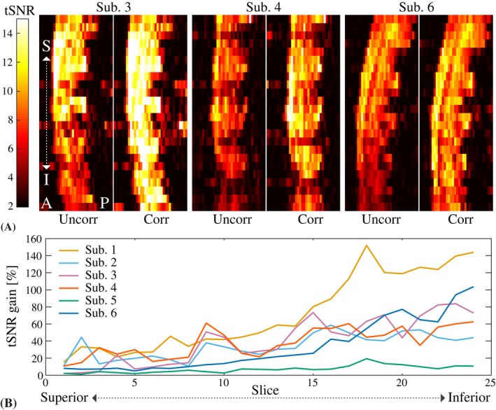
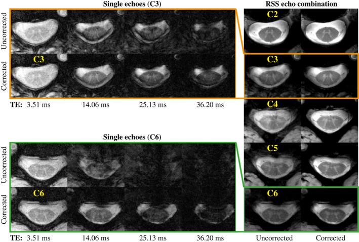
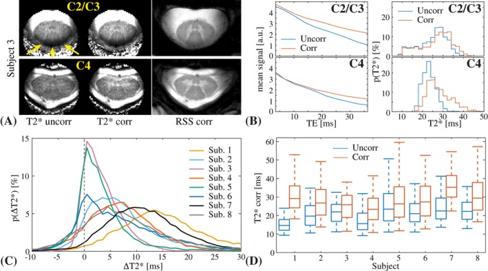
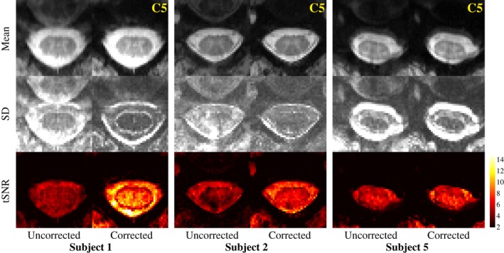
In the multi-echo acquisition, breathing-induced fields caused severe ghosting in images with long TE, which led to a systematic underestimation of T2* in the spinal cord. The trace-based correction reduced the ghosting and increased the estimated T2* values. Breathing-related ghosting was also observed in the multi-shot EPI images. The correction largely removed the ghosting, thereby improving the temporal signal-to-noise ratio of the time series.
Trace-based retrospective correction of breathing-induced field variations can reduce ghosting and improve quantitative metrics in multi-shot structural and functional T2*-weighted imaging of the spinal cord. The method is straightforward to implement and does not rely on sequence modifications or additional hardware beyond a respiratory bellows.
超高磁场下的脊髓 MRI 受到与呼吸周期相关的时变磁场的限制,导致多-shot 采集的鬼影伪影。在这里,我们提出了一种基于将呼吸波纹管的信号与脊髓内磁场变化相关联的校正方法。该信息用于在图像重建水平上校正数据。
在 7T 下对颈椎脊髓进行多-shot T2*-加权成像的背景下进行了校正。在高分辨率多回波梯度回波序列期间采集呼吸轨迹,用于结构成像和定量 T2*映射,以及多-shot EPI 时间序列,适用于 fMRI。在每个人中,通过短的校准扫描确定轨迹与呼吸诱导场之间的耦合。使用和不使用基于轨迹的校正重建图像。
在多回波采集期间,呼吸诱导的场在长 TE 的图像中引起严重的鬼影,这导致脊髓中 T2的系统低估。基于轨迹的校正减少了鬼影并增加了估计的 T2值。在多-shot EPI 图像中也观察到与呼吸相关的鬼影。校正大大消除了鬼影,从而提高了时间序列的时间信号到噪声比。
基于轨迹的呼吸诱导场变化的回溯校正可以减少鬼影并改善脊髓多-shot 结构和功能 T2*-加权成像的定量指标。该方法易于实现,不依赖于序列修改或除呼吸波纹管之外的额外硬件。