Estep Michael, Mehta Rohini, Bratthauer Gary, Alaparthi Lakshmi, Monge Fanny, Ali Simon, Abdelatif Dinan, Younoszai Zahra, Stepanova Maria, Goodman Zachary D, Younossi Zobair M
Center for Liver Diseases, Department of Medicine, Inova Fairfax Hospital, Virginia, USA.
Betty and Guy Beatty Center for Integrated Research, Claude Moore Health Education and Research Building, Inova Health System, 3300 Gallows Road, Falls Church, VA, 22042, USA.
BMC Gastroenterol. 2019 Feb 11;19(1):27. doi: 10.1186/s12876-019-0951-y.
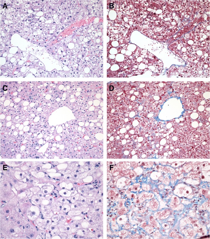
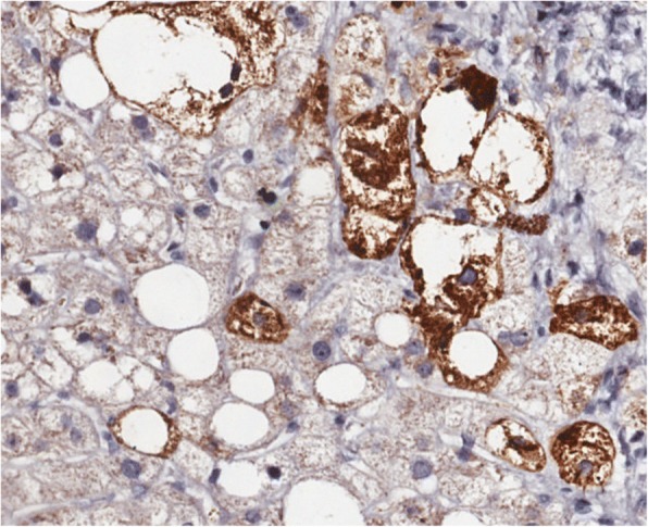
Hepatic expression of Sonic Hedgehog (SHH) is associated with Non-alcoholic fatty liver disease (NAFLD) and development of Non-alcoholic steatohepatitis (NASH). Hepatic SHH detection increases with the diagnosis of NASH. This pilot study was designed to confirm that staining for SHH is useful in NASH diagnosis and determine whether quantification of staining by computer assisted morphometry (CAM) can be used to assess severity of ballooning degeneration.
SHH was detected by immunohistochemistry (IHC) on paraffin-embedded liver sections in subjects (N = 69) with biopsy proven NAFLD and no liver disease (control). Serum samples were also available for these subjects. Post-staining, a digitized image of the section was acquired and an area quantification algorithm was used to quantify the degree of SHH expression. Additionally, circulating M30, M65, and SHH were measured by ELISA.
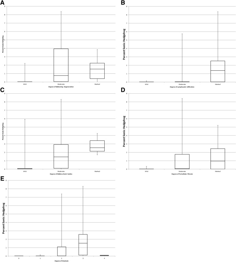
Notably, hepatic SHH expression correlated with histologic ballooning degeneration (rho = 0.62, p < 0.0001), steatosis grade (rho = 0.554, P < 0.001), Mallory-Denk bodies (rho = 0.54, P < 0.001), pericellular fibrosis (rho = 0.527, P < 0.001), and lymphocytic infiltration (rho = 0.435, P < 0.0002). Additionally, hepatic SHH expression correlated with circulating M65 (rho = 0.588, p < 0.0001), and circulating M30 (rho = 0.375, p = 0.001), as well as AST and ALT (rho = 0.43, p = 0.0004, and rho = 0.27, p = 0.03, respectively). Further, serum M30 was almost twice as high in NASH patients compared to non-NASH (539.1 ± 290.8 U/L vs. 287.6 ± 190.5 U/L; p = 0.0002), while M65 was almost three times higher in NASH patients compared to non-NASH (441.2 ± 464.2 U/L vs. 162.8 ± 353.1 U/L, P = 0.0006). Logistic modeling indicates hepatic SHH expression and presence of type 2 diabetes as independent predictors of advanced fibrosis (defined as portal and pericellular fibrosis > 2: OR = 1.986, p = 0.01, and OR = 3.280, p = 0.03, respectively).
Thus, our findings show quantitation of SHH expression by CAM can provide a tool for quantifying changes in hepatocyte injury and assist in unambiguous staging/grading of NASH. Our study showed minimal interobserver variability using CAM based quantification. Once validated, CAM assessment of hepatic SHH could benefit clinical trials or long term outcomes studies of NASH subjects.
音猬因子(SHH)的肝脏表达与非酒精性脂肪性肝病(NAFLD)以及非酒精性脂肪性肝炎(NASH)的发展相关。随着NASH的诊断,肝脏中SHH的检测率增加。这项初步研究旨在证实SHH染色对NASH诊断有用,并确定通过计算机辅助形态测量法(CAM)对染色进行定量是否可用于评估气球样变性的严重程度。
通过免疫组织化学(IHC)在经活检证实患有NAFLD且无肝脏疾病的受试者(N = 69)(对照组)的石蜡包埋肝切片上检测SHH。这些受试者也有血清样本。染色后,获取切片的数字化图像,并使用面积定量算法对SHH表达程度进行定量。此外,通过酶联免疫吸附测定(ELISA)测量循环中的M30、M65和SHH。
值得注意的是,肝脏SHH表达与组织学气球样变性(rho = 0.62,p < 0.0001)、脂肪变性分级(rho = 0.554,P < 0.001)、马洛里-登克小体(rho = 0.54,P < 0.001)、细胞周围纤维化(rho = 0.527,P < 0.001)和淋巴细胞浸润(rho = 0.435,P < 0.0002)相关。此外,肝脏SHH表达与循环中的M65(rho = 0.588,p < 0.0001)、循环中的M30(rho = 0.375,p = 0.001)以及天冬氨酸转氨酶(AST)和丙氨酸转氨酶(ALT)(分别为rho = 0.43,p = 0.0004和rho = 0.27,p = 0.03)相关。此外,与非NASH患者相比,NASH患者的血清M30几乎高出两倍(539.1±290.8 U/L对287.6±190.5 U/L;p = 0.0002),而与非NASH患者相比,NASH患者的M65几乎高出三倍(441.2±464.2 U/L对162.8±353.1 U/L,P = 0.0006)。逻辑模型表明肝脏SHH表达和2型糖尿病的存在是晚期纤维化(定义为门静脉和细胞周围纤维化> 2)的独立预测因素(分别为OR = 1.986,p = 0.01和OR = 3.280,p = 0.03)。
因此,我们的研究结果表明,通过CAM对SHH表达进行定量可为量化肝细胞损伤变化提供一种工具,并有助于对NASH进行明确的分期/分级。我们的研究表明,使用基于CAM的定量方法,观察者间的变异性最小。一旦得到验证,肝脏SHH的CAM评估可能会使NASH受试者的临床试验或长期结局研究受益。