• 文献检索
  • 文档翻译
  • 深度研究
  • 学术资讯
  • Suppr Zotero 插件Zotero 插件
  • 邀请有礼
  • 套餐&价格
  • 历史记录
应用&插件
Suppr Zotero 插件Zotero 插件浏览器插件Mac 客户端Windows 客户端微信小程序
定价
高级版会员购买积分包购买API积分包
服务
文献检索文档翻译深度研究API 文档MCP 服务
关于我们
关于 Suppr公司介绍联系我们用户协议隐私条款
关注我们

Suppr 超能文献

核心技术专利:CN118964589B侵权必究
粤ICP备2023148730 号-1Suppr @ 2026

文献检索

告别复杂PubMed语法,用中文像聊天一样搜索,搜遍4000万医学文献。AI智能推荐,让科研检索更轻松。

立即免费搜索

文件翻译

保留排版,准确专业,支持PDF/Word/PPT等文件格式,支持 12+语言互译。

免费翻译文档

深度研究

AI帮你快速写综述,25分钟生成高质量综述,智能提取关键信息,辅助科研写作。

立即免费体验

表皮生长因子受体在恶性浆液性和黏液性卵巢肿瘤中的免疫表达

EGFR Immunoexpression in Malignant Serous and Mucinous Ovarian Tumors.

作者信息

Cîrstea A E, Stepan A E, Zăvoi R E, Simionescu C E

机构信息

PhD Student, University of Medicine and Pharmacy of Craiova, Romania.

Department of Pathology, University of Medicine and Pharmacy of Craiova, Romania.

出版信息

Curr Health Sci J. 2018 Apr-Jun;44(2):129-134. doi: 10.12865/CHSJ.44.02.06. Epub 2018 Mar 27.

DOI:10.12865/CHSJ.44.02.06
PMID:30746159
原文链接:https://pmc.ncbi.nlm.nih.gov/articles/PMC6320459/
Abstract

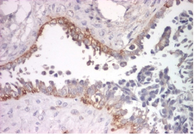
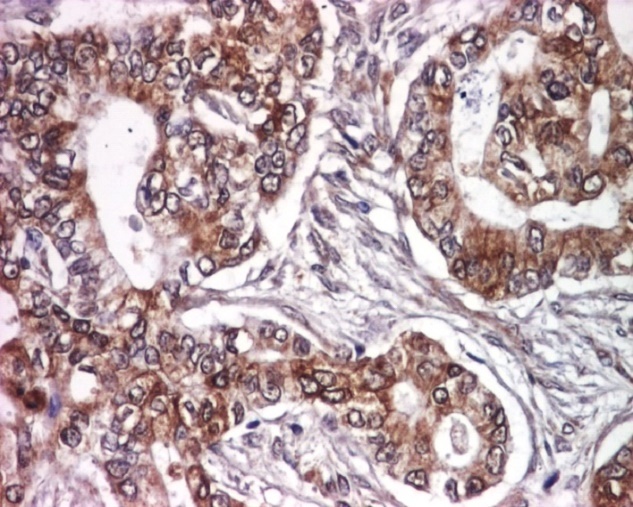
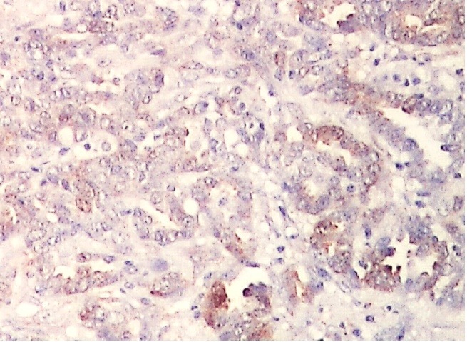
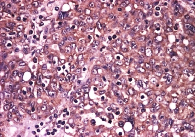
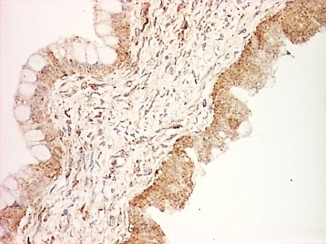
The epithelial growth factor receptor (EGFR) is involved in various stages of cancer growth such as tumor initiation, angiogenesis and metastasis, being an attractive target for oncogenic therapy. The present study aims to evaluate the EGFR expression in 54 cases of serous and mucinous ovarian borderline tumors and carcinomas. EGFR expression was present in more than half of the investigated tumors, more frequently in carcinomas than in borderline tumors, especially in the serous type. The highest values of the final staining score (FSS) were observed only in serous carcinomas in the advanced stages of the disease. As a result of frequent expression of EGFR in ovarian tumors, it is necessary to monitor EGFR inhibitors for ovarian cancer therapy, but probably after establishing more rigorous selection and stratification criteria for patients.

摘要

表皮生长因子受体(EGFR)参与癌症生长的各个阶段,如肿瘤起始、血管生成和转移,是肿瘤治疗的一个有吸引力的靶点。本研究旨在评估54例浆液性和黏液性卵巢交界性肿瘤及癌中EGFR的表达情况。超过半数的被研究肿瘤中存在EGFR表达,癌中的表达比交界性肿瘤更常见,尤其是浆液性类型。仅在疾病晚期的浆液性癌中观察到最终染色评分(FSS)的最高值。由于EGFR在卵巢肿瘤中频繁表达,有必要监测EGFR抑制剂用于卵巢癌治疗,但可能需要先为患者建立更严格的选择和分层标准。

https://cdn.ncbi.nlm.nih.gov/pmc/blobs/e6ce/6320459/84f9613663cc/CHSJ-44-2-129-fig5.jpg
https://cdn.ncbi.nlm.nih.gov/pmc/blobs/e6ce/6320459/c10379a6ddcf/CHSJ-44-2-129-fig1.jpg
https://cdn.ncbi.nlm.nih.gov/pmc/blobs/e6ce/6320459/4a7125ae1a6d/CHSJ-44-2-129-fig2.jpg
https://cdn.ncbi.nlm.nih.gov/pmc/blobs/e6ce/6320459/687e7cd3774e/CHSJ-44-2-129-fig3.jpg
https://cdn.ncbi.nlm.nih.gov/pmc/blobs/e6ce/6320459/f9b4d01f1aea/CHSJ-44-2-129-fig4.jpg
https://cdn.ncbi.nlm.nih.gov/pmc/blobs/e6ce/6320459/84f9613663cc/CHSJ-44-2-129-fig5.jpg

相似文献

1
EGFR Immunoexpression in Malignant Serous and Mucinous Ovarian Tumors.表皮生长因子受体在恶性浆液性和黏液性卵巢肿瘤中的免疫表达
Curr Health Sci J. 2018 Apr-Jun;44(2):129-134. doi: 10.12865/CHSJ.44.02.06. Epub 2018 Mar 27.
2
The immunoexpression of EGFR, HER2 and HER3 in malignant serous ovarian tumors.表皮生长因子受体(EGFR)、人表皮生长因子受体2(HER2)和人表皮生长因子受体3(HER3)在恶性浆液性卵巢肿瘤中的免疫表达
Rom J Morphol Embryol. 2017;58(4):1269-1273.
3
Clinical factors and biomarkers in ovarian tumors development.卵巢肿瘤发生发展中的临床因素和生物标志物
Rom J Morphol Embryol. 2008;49(3):327-38.
4
Expression of cell regulatory proteins in ovarian borderline tumors.
Cancer. 1996 May 15;77(10):2092-8. doi: 10.1002/(SICI)1097-0142(19960515)77:10<2092::AID-CNCR19>3.0.CO;2-Q.
5
Significance of cell cycle regulatory proteins as malignant and prognostic biomarkers in ovarian epithelial tumors.细胞周期调控蛋白作为卵巢上皮性肿瘤恶性和预后生物标志物的意义。
Int J Gynecol Pathol. 2011 May;30(3):205-17. doi: 10.1097/PGP.0b013e3182063e71.
6
Phosphorylated 4E binding protein 1: a hallmark of cell signaling that correlates with survival in ovarian cancer.磷酸化4E结合蛋白1:一种与卵巢癌生存相关的细胞信号传导标志。
Cancer. 2006 Oct 15;107(8):1801-11. doi: 10.1002/cncr.22195.
7
Detection of HER-2 and EGFR gene amplification using chromogenic in-situ hybridization technique in ovarian tumors.采用显色原位杂交技术检测卵巢肿瘤中HER-2和EGFR基因扩增情况。
Appl Immunohistochem Mol Morphol. 2010 Jan;18(1):69-74. doi: 10.1097/PAI.0b013e3181af7d3f.
8
The immunohistochemical expression of p53 and Ki67 in ovarian epithelial borderline tumors. Correlation with clinicopathological factors.p53和Ki67在卵巢上皮性交界性肿瘤中的免疫组化表达。与临床病理因素的相关性。
Rom J Morphol Embryol. 2012;53(4):967-73.
9
[Clinicopathologic analysis and expression of cyclin D1 and p53 of ovarian borderline tumors and carcinomas].卵巢交界性肿瘤和癌的临床病理分析及细胞周期蛋白D1和p53的表达
Zhonghua Fu Chan Ke Za Zhi. 2007 Apr;42(4):227-32.
10
Podocalyxin-like protein 1 expression and correlation with clinical characteristics in epithelial serous and mucinous ovarian carcinoma and tumor-like lesions.足细胞蛋白样蛋白 1 在卵巢上皮性浆液性和黏液性癌及肿瘤样病变中的表达及其与临床特征的相关性。
Pathobiology. 2012;79(6):307-13. doi: 10.1159/000338078. Epub 2012 Jun 9.

引用本文的文献

1
Epidermal growth factor receptor ligands enriched in follicular fluid exosomes promote oncogenesis of fallopian tube epithelial cells.富含卵泡液外泌体的表皮生长因子受体配体促进输卵管上皮细胞的肿瘤发生。
Cancer Cell Int. 2024 Dec 21;24(1):424. doi: 10.1186/s12935-024-03614-9.
2
The Protein Landscape of Mucinous Ovarian Cancer: Towards a Theranostic.黏液性卵巢癌的蛋白质图谱:迈向诊疗一体化
Cancers (Basel). 2021 Nov 9;13(22):5596. doi: 10.3390/cancers13225596.
3
Genome-wide DNA copy number profiling and bioinformatics analysis of ovarian cancer reveals key genes and pathways associated with distinct invasive/migratory capabilities.

本文引用的文献

1
EGFR as a prognostic biomarker and therapeutic target in ovarian cancer: evaluation of patient cohort and literature review.表皮生长因子受体作为卵巢癌的预后生物标志物和治疗靶点:患者队列评估及文献综述
Genes Cancer. 2017 May;8(5-6):589-599. doi: 10.18632/genesandcancer.142.
2
Increased serum level of epidermal growth factor receptor (EGFR) is associated with poor progression-free survival in patients with epithelial ovarian cancer.血清表皮生长因子受体(EGFR)水平升高与上皮性卵巢癌患者无进展生存期不良相关。
Cancer Chemother Pharmacol. 2014 Mar;73(3):631-7. doi: 10.1007/s00280-014-2396-x. Epub 2014 Jan 30.
3
Overexpression of HIF-1α, metallothionein and SLUG is associated with high TNM stage and lymph node metastasis in papillary thyroid carcinoma.
卵巢癌全基因组 DNA 拷贝数谱分析和生物信息学分析揭示了与不同侵袭/迁移能力相关的关键基因和途径。
Aging (Albany NY). 2020 Jan 2;12(1):178-192. doi: 10.18632/aging.102608.
4
Lidocaine inhibits the progression of retinoblastoma in vitro and in vivo by modulating the miR‑520a‑3p/EGFR axis.利多卡因通过调节 miR-520a-3p/EGFR 轴抑制视网膜母细胞瘤的体外和体内进展。
Mol Med Rep. 2019 Aug;20(2):1333-1342. doi: 10.3892/mmr.2019.10363. Epub 2019 Jun 6.
缺氧诱导因子-1α、金属硫蛋白和SLUG的过表达与甲状腺乳头状癌的高TNM分期及淋巴结转移相关。
Int J Clin Exp Pathol. 2013 Dec 15;7(1):322-30. eCollection 2014.
4
Comprehensive profiling of EGFR/HER receptors for personalized treatment of gynecologic cancers.EGFR/HER 受体的综合分析用于妇科癌症的个体化治疗。
Mol Diagn Ther. 2014 Apr;18(2):137-51. doi: 10.1007/s40291-013-0070-3.
5
Effect of BRCA1 on epidermal growth factor receptor in ovarian cancer.BRCA1对卵巢癌中表皮生长因子受体的影响。
J Exp Clin Cancer Res. 2013 Dec 9;32(1):102. doi: 10.1186/1756-9966-32-102.
6
Epidermal growth factor receptor targeting in cancer: a review of trends and strategies.表皮生长因子受体靶向治疗在癌症中的应用:趋势与策略综述。
Biomaterials. 2013 Nov;34(34):8690-707. doi: 10.1016/j.biomaterials.2013.07.100. Epub 2013 Aug 13.
7
Targeting the ERBB family in cancer: couples therapy.针对癌症中的 ERBB 家族:夫妻治疗。
Nat Rev Cancer. 2013 Sep;13(9):663-73. doi: 10.1038/nrc3559. Epub 2013 Aug 16.
8
Profile of erlotinib and its potential in the treatment of advanced ovarian carcinoma.厄洛替尼的特性及其在治疗晚期卵巢癌中的潜力。
Onco Targets Ther. 2013 Apr 18;6:427-35. doi: 10.2147/OTT.S30373. Print 2013.
9
GPR30 regulates the EGFR-Akt cascade and predicts lower survival in patients with ovarian cancer.GPR30 调节 EGFR-Akt 级联反应,并预测卵巢癌患者的生存率降低。
J Ovarian Res. 2012 Nov 19;5(1):35. doi: 10.1186/1757-2215-5-35.
10
EGFR, HER2÷neu and Ki67 immunoexpression in serous ovarian tumors.浆液性卵巢肿瘤中表皮生长因子受体(EGFR)、人表皮生长因子受体2(HER2÷neu)及Ki67免疫表达情况
Rom J Morphol Embryol. 2012;53(3):563-7.