• 文献检索
  • 文档翻译
  • 深度研究
  • 学术资讯
  • Suppr Zotero 插件Zotero 插件
  • 邀请有礼
  • 套餐&价格
  • 历史记录
应用&插件
Suppr Zotero 插件Zotero 插件浏览器插件Mac 客户端Windows 客户端微信小程序
定价
高级版会员购买积分包购买API积分包
服务
文献检索文档翻译深度研究API 文档MCP 服务
关于我们
关于 Suppr公司介绍联系我们用户协议隐私条款
关注我们

Suppr 超能文献

核心技术专利:CN118964589B侵权必究
粤ICP备2023148730 号-1Suppr @ 2026

文献检索

告别复杂PubMed语法,用中文像聊天一样搜索,搜遍4000万医学文献。AI智能推荐,让科研检索更轻松。

立即免费搜索

文件翻译

保留排版,准确专业,支持PDF/Word/PPT等文件格式,支持 12+语言互译。

免费翻译文档

深度研究

AI帮你快速写综述,25分钟生成高质量综述,智能提取关键信息,辅助科研写作。

立即免费体验

上皮-间充质转化标志物和 CD44 异构体在前列腺癌细胞的 2D 和 3D 细胞培养中表达不同。

Epithelial-To-Mesenchymal Transition Markers and CD44 Isoforms Are Differently Expressed in 2D and 3D Cell Cultures of Prostate Cancer Cells.

机构信息

Department of Excellence: Department of Pharmacological and Biomolecular Sciences, Università degli Studi di Milano, via Balzaretti 9, 20133 Milan, Italy.

Department of Biomedical Sciences for Health, Università degli Studi di Milano, via Mangiagalli 31, 20133 Milan, Italy.

出版信息

Cells. 2019 Feb 11;8(2):143. doi: 10.3390/cells8020143.

DOI:10.3390/cells8020143
PMID:30754655
原文链接:https://pmc.ncbi.nlm.nih.gov/articles/PMC6406374/
Abstract

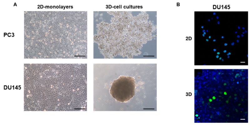
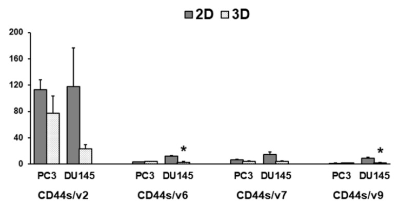
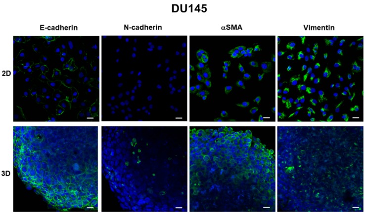
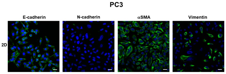
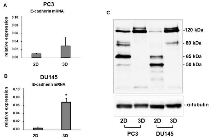
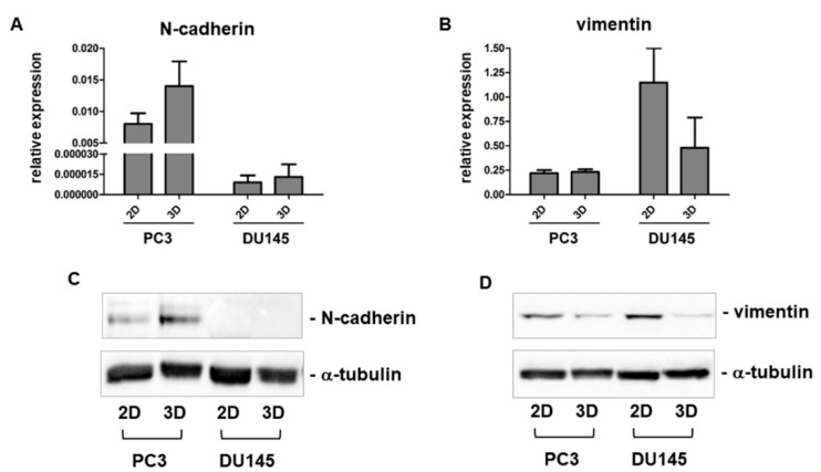
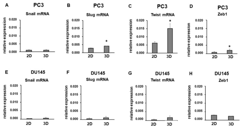
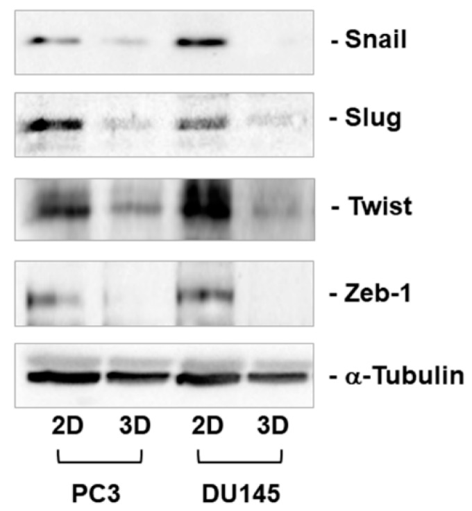
Three-dimensional (3D) cell cultures allow the mimic of functions of living tissues andprovide key information encoded in tissue architecture. Considered the pivotal role of epithelial-tomesenchymaltransition (EMT) in carcinoma progression, including prostate cancer (PCa), weaimed at investigating the effect of the 3D arrangement on the expression of some key markers ofEMT in cultured human prostate cancer (PCa) cells, to better understand PCa cell behavior. PC3 andDU145 PCa cells were cultured in RPMI cell culture medium either in 2D-monolayers or in 3Dspheroids.The main EMT markers E-cadherin, N-cadherin, α-smooth muscle actin (αSMA),vimentin, Snail, Slug, Twist and Zeb1 were evaluated by confocal microscopy, real-time PCR andWestern blot. Confocal microscopy revealed that E-cadherin was similarly expressed at the cellboundaries on the plasma membrane of PCa cells grown in 2D-monolayers, as well as in 3Dspheroids,but resulted up-regulated in 3D-spheroids, compared to 2D-monolayers, at the mRNAand protein level. Moreover, markers of the mesenchymal phenotype were expressed at very lowlevels in 3D-spheroids, suggesting important differences in the phenotype of PCa cells grown in 3Dspheroidsor in 2D-monolayers. Considered as a whole, our findings contribute to a clarification ofthe role of EMT in PCa and confirm that a 3D cell culture model could provide deeper insight intothe understanding of the biology of PCa.

摘要

三维(3D)细胞培养允许模拟活组织的功能,并提供组织架构中编码的关键信息。考虑到上皮-间充质转化(EMT)在包括前列腺癌(PCa)在内的癌进展中的关键作用,我们旨在研究 3D 排列对培养的人前列腺癌(PCa)细胞中一些 EMT 关键标志物表达的影响,以更好地理解 PCa 细胞的行为。PC3 和 DU145 PCa 细胞在 RPMI 细胞培养基中分别在 2D 单层或 3D 球体中培养。通过共聚焦显微镜、实时 PCR 和 Western blot 评估 EMT 的主要标志物 E-钙粘蛋白、N-钙粘蛋白、α-平滑肌肌动蛋白(αSMA)、波形蛋白、Snail、Slug、Twist 和 Zeb1。共聚焦显微镜显示,E-钙粘蛋白在 2D 单层培养的 PCa 细胞的质膜上的细胞边界处同样表达,并且在 3D 球体中也表达,但与 2D 单层相比,在 mRNA 和蛋白水平上均上调。此外,间充质表型标志物在 3D 球体中表达水平非常低,表明在 3D 球体中培养的 PCa 细胞与在 2D 单层中培养的 PCa 细胞的表型存在重要差异。总的来说,我们的发现有助于阐明 EMT 在 PCa 中的作用,并证实 3D 细胞培养模型可以更深入地了解 PCa 的生物学。

https://cdn.ncbi.nlm.nih.gov/pmc/blobs/3f15/6406374/7d0d05f2c5ef/cells-08-00143-g009.jpg
https://cdn.ncbi.nlm.nih.gov/pmc/blobs/3f15/6406374/bf962b0a30db/cells-08-00143-g001.jpg
https://cdn.ncbi.nlm.nih.gov/pmc/blobs/3f15/6406374/12c206dc4f05/cells-08-00143-g002.jpg
https://cdn.ncbi.nlm.nih.gov/pmc/blobs/3f15/6406374/f2854beb789e/cells-08-00143-g003.jpg
https://cdn.ncbi.nlm.nih.gov/pmc/blobs/3f15/6406374/aa6fc4031685/cells-08-00143-g004.jpg
https://cdn.ncbi.nlm.nih.gov/pmc/blobs/3f15/6406374/74293d058743/cells-08-00143-g005.jpg
https://cdn.ncbi.nlm.nih.gov/pmc/blobs/3f15/6406374/1276bf9b9937/cells-08-00143-g006.jpg
https://cdn.ncbi.nlm.nih.gov/pmc/blobs/3f15/6406374/b0e334f029f0/cells-08-00143-g007.jpg
https://cdn.ncbi.nlm.nih.gov/pmc/blobs/3f15/6406374/d4ef0f896ca6/cells-08-00143-g008.jpg
https://cdn.ncbi.nlm.nih.gov/pmc/blobs/3f15/6406374/7d0d05f2c5ef/cells-08-00143-g009.jpg

相似文献

1
Epithelial-To-Mesenchymal Transition Markers and CD44 Isoforms Are Differently Expressed in 2D and 3D Cell Cultures of Prostate Cancer Cells.上皮-间充质转化标志物和 CD44 异构体在前列腺癌细胞的 2D 和 3D 细胞培养中表达不同。
Cells. 2019 Feb 11;8(2):143. doi: 10.3390/cells8020143.
2
Epithelial-to-mesenchymal transition in pancreatic ductal adenocarcinoma: Characterization in a 3D-cell culture model.胰腺导管腺癌中的上皮-间质转化:三维细胞培养模型中的特征
World J Gastroenterol. 2016 May 14;22(18):4466-83. doi: 10.3748/wjg.v22.i18.4466.
3
Bone-stromal cells up-regulate tumourigenic markers in a tumour-stromal 3D model of prostate cancer.成骨基质细胞在前列腺癌的肿瘤基质 3D 模型中上调致瘤标志物。
Mol Cancer. 2013 Sep 30;12(1):112. doi: 10.1186/1476-4598-12-112.
4
Differentiation States of Phenotypic Transition of Melanoma Cells Are Revealed by 3D Cell Cultures.三维细胞培养揭示了黑色素瘤细胞表型转化的分化状态。
Cells. 2024 Jan 17;13(2):181. doi: 10.3390/cells13020181.
5
Down-regulation of E-cadherin enhances prostate cancer chemoresistance via Notch signaling.E-钙黏蛋白的下调通过Notch信号通路增强前列腺癌的化疗耐药性。
Chin J Cancer. 2017 Mar 29;36(1):35. doi: 10.1186/s40880-017-0203-x.
6
Comprehensive study of gene and microRNA expression related to epithelial-mesenchymal transition in prostate cancer.前列腺癌中与上皮-间质转化相关的基因和微小RNA表达的综合研究。
PLoS One. 2014 Nov 19;9(11):e113700. doi: 10.1371/journal.pone.0113700. eCollection 2014.
7
Rosuvastatin inhibit spheroid formation and epithelial-mesenchymal transition (EMT) in prostate cancer PC-3 cell line.罗苏伐他汀抑制前列腺癌细胞系 PC-3 球体形成和上皮-间充质转化(EMT)。
Mol Biol Rep. 2020 Nov;47(11):8727-8737. doi: 10.1007/s11033-020-05918-1. Epub 2020 Oct 21.
8
In vivo biomarker expression patterns are preserved in 3D cultures of Prostate Cancer.在前列腺癌的 3D 培养物中,生物标志物的表达模式得以保持。
Exp Cell Res. 2012 Nov 15;318(19):2507-19. doi: 10.1016/j.yexcr.2012.07.013. Epub 2012 Jul 27.
9
Skp2 and Slug Are Coexpressed in Aggressive Prostate Cancer and Inhibited by Neddylation Blockade.Skp2 和 Slug 在侵袭性前列腺癌中共同表达,并受泛素化阻断抑制。
Int J Mol Sci. 2021 Mar 11;22(6):2844. doi: 10.3390/ijms22062844.
10
TGF-β1 promotes epithelial-to-mesenchymal transition and stemness of prostate cancer cells by inducing PCBP1 degradation and alternative splicing of CD44.TGF-β1 通过诱导 PCBP1 降解和 CD44 的可变剪接促进前列腺癌细胞的上皮间质转化和干细胞特性。
Cell Mol Life Sci. 2021 Feb;78(3):949-962. doi: 10.1007/s00018-020-03544-5. Epub 2020 May 21.

引用本文的文献

1
Advances in research regarding epithelial-mesenchymal transition and prostate cancer.上皮-间质转化与前列腺癌的研究进展
Front Cell Dev Biol. 2025 May 30;13:1583255. doi: 10.3389/fcell.2025.1583255. eCollection 2025.
2
Mimicking the Complexity of Solid Tumors: How Spheroids Could Advance Cancer Preclinical Transformative Approaches.模拟实体瘤的复杂性:球体如何推动癌症临床前变革性方法的发展。
Cancers (Basel). 2025 Mar 30;17(7):1161. doi: 10.3390/cancers17071161.
3
Patient-Derived Breast Cancer Bone Metastasis Model Using Bone-Mimetic Nanoclay Scaffolds.

本文引用的文献

1
Epithelial-Mesenchymal Transition (EMT) and Prostate Cancer.上皮-间充质转化(EMT)与前列腺癌。
Adv Exp Med Biol. 2018;1095:101-110. doi: 10.1007/978-3-319-95693-0_6.
2
Global cancer statistics 2018: GLOBOCAN estimates of incidence and mortality worldwide for 36 cancers in 185 countries.全球癌症统计数据 2018:GLOBOCAN 对全球 185 个国家/地区 36 种癌症的发病率和死亡率的估计。
CA Cancer J Clin. 2018 Nov;68(6):394-424. doi: 10.3322/caac.21492. Epub 2018 Sep 12.
3
Interleukin-4 induces a CD44 /CD49b PC3 subpopulation with tumor-initiating characteristics.
使用仿骨纳米粘土支架构建的患者来源的乳腺癌骨转移模型
J Tissue Eng Regen Med. 2023 Mar 11;2023:5753666. doi: 10.1155/2023/5753666. eCollection 2023.
4
Detection of circulating tumor cells in non-metastatic prostate cancer through integration of a microfluidic CTC enrichment system and multiparametric flow cytometry.通过微流控 CTC 富集系统和多参数流式细胞术检测非转移性前列腺癌中的循环肿瘤细胞。
PLoS One. 2024 Oct 23;19(10):e0312296. doi: 10.1371/journal.pone.0312296. eCollection 2024.
5
The biological function of extracellular vesicles in prostate cancer and their clinical application as diagnostic and prognostic biomarkers.细胞外囊泡在前列腺癌中的生物学功能及其作为诊断和预后生物标志物的临床应用。
Cancer Metastasis Rev. 2024 Dec;43(4):1611-1627. doi: 10.1007/s10555-024-10210-w. Epub 2024 Sep 24.
6
Long-term zinc treatment alters the mechanical properties and metabolism of prostate cancer cells.长期锌治疗会改变前列腺癌细胞的力学性能和代谢。
Cancer Cell Int. 2024 Sep 11;24(1):313. doi: 10.1186/s12935-024-03495-y.
7
The Pros and Cons of Estrogens in Prostate Cancer: An Update with a Focus on Phytoestrogens.雌激素在前列腺癌中的利弊:聚焦植物雌激素的最新进展
Biomedicines. 2024 Jul 23;12(8):1636. doi: 10.3390/biomedicines12081636.
8
Liver X Receptors Enhance Epithelial to Mesenchymal Transition in Metastatic Prostate Cancer Cells.肝脏X受体增强转移性前列腺癌细胞的上皮-间质转化
Cancers (Basel). 2024 Aug 6;16(16):2776. doi: 10.3390/cancers16162776.
9
Differentiation States of Phenotypic Transition of Melanoma Cells Are Revealed by 3D Cell Cultures.三维细胞培养揭示了黑色素瘤细胞表型转化的分化状态。
Cells. 2024 Jan 17;13(2):181. doi: 10.3390/cells13020181.
10
Scaffold-based 3D cell culture models in cancer research.基于支架的 3D 细胞培养模型在癌症研究中的应用。
J Biomed Sci. 2024 Jan 14;31(1):7. doi: 10.1186/s12929-024-00994-y.
白细胞介素-4 诱导具有肿瘤起始特性的 CD44/CD49b PC3 亚群。
J Cell Biochem. 2018 May;119(5):4103-4112. doi: 10.1002/jcb.26607. Epub 2018 Jan 19.
4
3D-spheroids: What can they tell us about pancreatic ductal adenocarcinoma cell phenotype?三维球体:它们能告诉我们关于胰腺导管腺癌细胞表型的哪些信息?
Exp Cell Res. 2017 Aug 15;357(2):299-309. doi: 10.1016/j.yexcr.2017.05.027. Epub 2017 May 29.
5
Sialylation facilitates self-assembly of 3D multicellular prostaspheres by using cyclo-RGDfK(TPP) peptide.唾液酸化通过使用环RGDfK(TPP)肽促进三维多细胞前列腺球的自组装。
Onco Targets Ther. 2017 May 4;10:2427-2447. doi: 10.2147/OTT.S133563. eCollection 2017.
6
Clinical significance of epithelial-mesenchymal transition markers in prostate cancer.上皮-间质转化标志物在前列腺癌中的临床意义
Hum Pathol. 2017 Mar;61:26-32. doi: 10.1016/j.humpath.2016.10.013. Epub 2016 Nov 4.
7
Epithelial-to-mesenchymal transition in pancreatic ductal adenocarcinoma: Characterization in a 3D-cell culture model.胰腺导管腺癌中的上皮-间质转化:三维细胞培养模型中的特征
World J Gastroenterol. 2016 May 14;22(18):4466-83. doi: 10.3748/wjg.v22.i18.4466.
8
The role of CD44 in epithelial-mesenchymal transition and cancer development.CD44在上皮-间质转化及癌症发展中的作用。
Onco Targets Ther. 2015 Dec 16;8:3783-92. doi: 10.2147/OTT.S95470. eCollection 2015.
9
Expression of α5-integrin, α7-integrin, Ε-cadherin, and N-cadherin in localized prostate cancer.α5整合素、α7整合素、E-钙黏蛋白和N-钙黏蛋白在局限性前列腺癌中的表达
Urol Oncol. 2016 Apr;34(4):165.e11-8. doi: 10.1016/j.urolonc.2015.10.016. Epub 2015 Dec 2.
10
N-cadherin promotes epithelial-mesenchymal transition and cancer stem cell-like traits via ErbB signaling in prostate cancer cells.N-钙黏蛋白通过ErbB信号通路促进前列腺癌细胞的上皮-间质转化和癌症干细胞样特征。
Int J Oncol. 2016 Feb;48(2):595-606. doi: 10.3892/ijo.2015.3270. Epub 2015 Nov 26.