Hollis Christin P, Dozier Alan K, Knutson Barbara L, Li Tonglei
Department of Pharmaceutical Sciences, University of Kentucky, Lexington, KY 40506, USA.
Electron Microscopy Center, University of Kentucky, Lexington, KY 40506, USA.
Acta Pharm Sin B. 2019 Jan;9(1):128-134. doi: 10.1016/j.apsb.2018.03.005. Epub 2018 Mar 22.
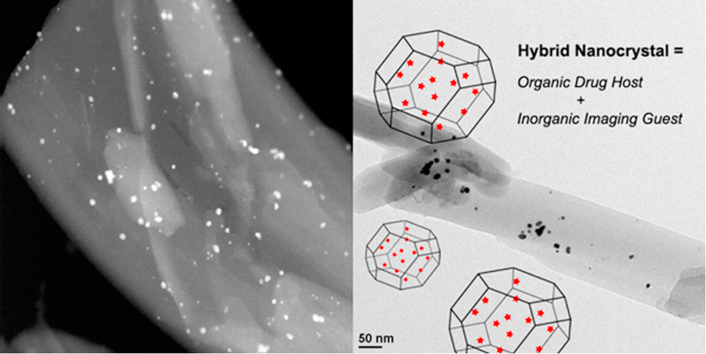
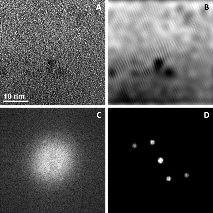
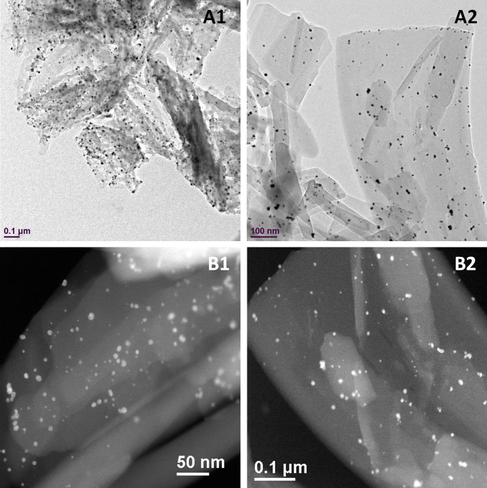
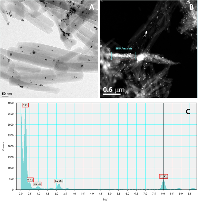
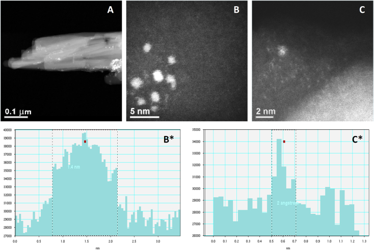
We demonstrate a novel inorganic-organic crystalline nanoconstruct, where gold atoms were imbedded in the crystal lattices as defects of camptothecin nanocrystals, suggesting its potential use as simultaneous agents for cancer therapy and bioimaging. The incorporation of gold, a potential computed tomography (CT) contrast agent, in the nanocrystals of camptothecin was detected by transmission electron microscope (TEM) and further quantified by energy dispersive X-ray spectrometry (EDS) and inductively coupled plasma-optical emission spectrometers (ICP-OES). Due to gold's high attenuation coefficient, only a relatively small amount needs to be present in order to create a good noise-to-contrast ratio in CT imaging. The imbedded gold atoms and clusters are expected to share the same biological fate as the camptothecin nanocrystals, reaching and accumulating in tumor site due to the enhanced permeation and retention (EPR) effect.
我们展示了一种新型的无机-有机晶体纳米结构,其中金原子作为喜树碱纳米晶体的缺陷嵌入晶格中,这表明其有潜力用作癌症治疗和生物成像的同步剂。通过透射电子显微镜(TEM)检测到喜树碱纳米晶体中掺入了具有潜在计算机断层扫描(CT)造影剂作用的金,并通过能量色散X射线光谱仪(EDS)和电感耦合等离子体发射光谱仪(ICP-OES)进一步定量。由于金的高衰减系数,在CT成像中仅需存在相对少量的金就能产生良好的信噪比。预计嵌入的金原子和团簇与喜树碱纳米晶体具有相同的生物学命运,由于增强的渗透和滞留(EPR)效应而到达并积聚在肿瘤部位。