Department of Orthodontics, Shanghai Stomatological Hospital, 1258 Fuxing Rd, 2nd Floor, Shanghai, 200000, China.
Department of Orthodontics, Ninth People's Hospital, College of Stomatology, Shanghai Jiao Tong University School of Medicine, 500 Quxi Rd, 7th Floor, Shanghai, 200000, China.
Prog Orthod. 2019 Feb 18;20(1):7. doi: 10.1186/s40510-019-0259-z.
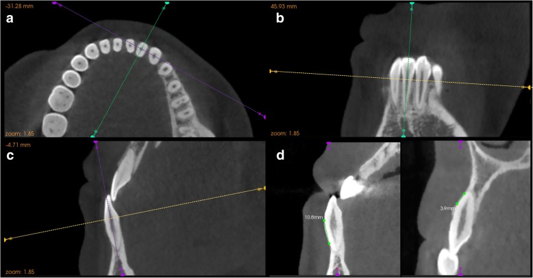
To evaluate the changes of alveolar dehiscence and fenestration after augmented corticotomy-assisted orthodontic treatment on cone-beam computed tomography (CBCT) compared with traditional pre-surgical orthodontics, both quantitatively and qualitatively.
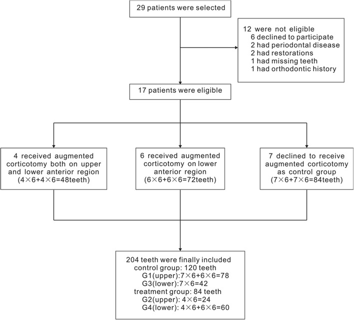
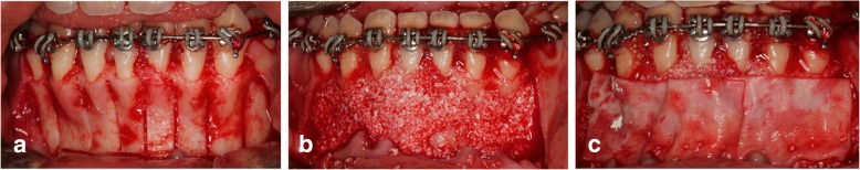
Two hundred and four anterior teeth from 17 skeletal class III malocclusions were divided into four groups. Groups G1 (upper teeth) and G3 (lower teeth), comprising 120 teeth, accepted traditional pre-surgical orthodontics; groups G2 (upper teeth) and G4(lower teeth), comprising 84 teeth, accepted augmented corticotomy-assisted pre-surgical orthodontics. The changes of alveolar bone dehiscence and fenestration of each tooth in all groups were evaluated with the help of CBCT.
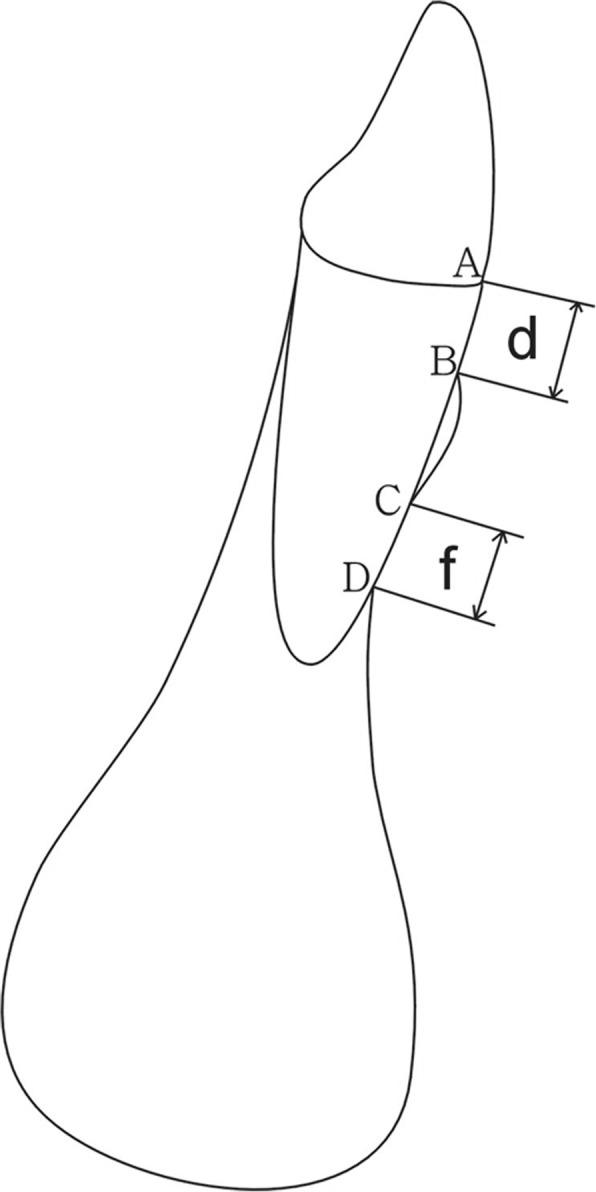
Quantitative analysis for comparing both groups: For the upper teeth, d - d was different between both groups while f - f was not statistically different. For the lower teeth, d - d was statistically different between both groups while f - f was not statistically different. Qualitative analysis: For the teeth that had no dehiscence before treatment, G2 and G4 had a better transition than did G1 and G3. For those having dehiscence before treatment, G4 had a better transition than did G3. For teeth having no fenestration before treatment, there was no statistically significant difference in transition between the control and treatment groups. For those having fenestration before treatment, G4 had a better transition than did G3.
For skeletal class III patients, augmented corticotomy-assisted orthodontic treatment is a promising method of improving alveolar bone dehiscence and fenestration for lower anterior teeth, and it also has the potential to protect both lower and upper anterior teeth against dehiscence.
通过锥形束 CT(CBCT)评估增强皮质切开辅助正畸治疗与传统术前正畸治疗后牙槽骨开窗和裂隙的变化,从定量和定性两方面进行评估。
17 例骨骼 III 类错畸形的 204 颗前牙分为 4 组。G1(上颌)和 G3(下颌)组共 120 颗牙接受传统术前正畸治疗;G2(上颌)和 G4(下颌)组共 84 颗牙接受增强皮质切开辅助术前正畸治疗。所有组的每颗牙的牙槽骨开窗和裂隙的变化均借助 CBCT 进行评估。
两组间定量分析比较:上颌牙,d-d 在两组间有差异,而 f-f 无统计学差异。下颌牙,d-d 在两组间有统计学差异,而 f-f 无统计学差异。定性分析:治疗前无开窗的牙齿,G2 和 G4 的过渡优于 G1 和 G3。治疗前有开窗的牙齿,G4 的过渡优于 G3。治疗前无裂隙的牙齿,对照组和治疗组之间的过渡无统计学差异。治疗前有裂隙的牙齿,G4 的过渡优于 G3。
对于骨骼 III 类患者,增强皮质切开辅助正畸治疗是改善下颌前牙牙槽骨开窗和裂隙的一种很有前途的方法,它还有可能保护下颌和上颌前牙免受开窗的影响。