• 文献检索
  • 文档翻译
  • 深度研究
  • 学术资讯
  • Suppr Zotero 插件Zotero 插件
  • 邀请有礼
  • 套餐&价格
  • 历史记录
应用&插件
Suppr Zotero 插件Zotero 插件浏览器插件Mac 客户端Windows 客户端微信小程序
定价
高级版会员购买积分包购买API积分包
服务
文献检索文档翻译深度研究API 文档MCP 服务
关于我们
关于 Suppr公司介绍联系我们用户协议隐私条款
关注我们

Suppr 超能文献

核心技术专利:CN118964589B侵权必究
粤ICP备2023148730 号-1Suppr @ 2026

文献检索

告别复杂PubMed语法,用中文像聊天一样搜索,搜遍4000万医学文献。AI智能推荐,让科研检索更轻松。

立即免费搜索

文件翻译

保留排版,准确专业,支持PDF/Word/PPT等文件格式,支持 12+语言互译。

免费翻译文档

深度研究

AI帮你快速写综述,25分钟生成高质量综述,智能提取关键信息,辅助科研写作。

立即免费体验

胸腺上皮肿瘤和胸腺囊肿的生长:放射学差异要点。

Growth of thymic epithelial tumors and thymic cysts: Differential radiological points.

机构信息

Department of Radiology, Seoul National University College of Medicine, and Institute of Radiation Medicine, Seoul National University Medical Research Center, Seoul, South Korea.

Cancer Research Institute, Seoul National University, Seoul, South Korea.

出版信息

Thorac Cancer. 2019 Apr;10(4):864-871. doi: 10.1111/1759-7714.13016. Epub 2019 Feb 21.

DOI:10.1111/1759-7714.13016
PMID:30793538
原文链接:https://pmc.ncbi.nlm.nih.gov/articles/PMC6449257/
Abstract

BACKGROUND

The growth rate of thymic epithelial tumors (TETs) and thymic cysts was investigated to determine whether they can be differentiated and clinico-radiological predictors of interval growth was identified.

METHODS

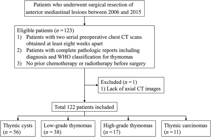
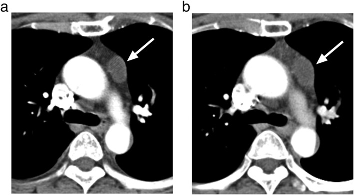
This retrospective study included 122 patients with pathologically proven thymic cysts (n = 56) or TETs (n = 66) who underwent two serial chest computed tomography scans at least eight weeks apart. Average diameters and attenuation were measured, volume-doubling times (VDTs) were calculated, and clinical characteristics were recorded. VDTs were compared using the log-rank test. Predictors of growth were analyzed using the log-rank test and Cox regression analysis.

RESULTS

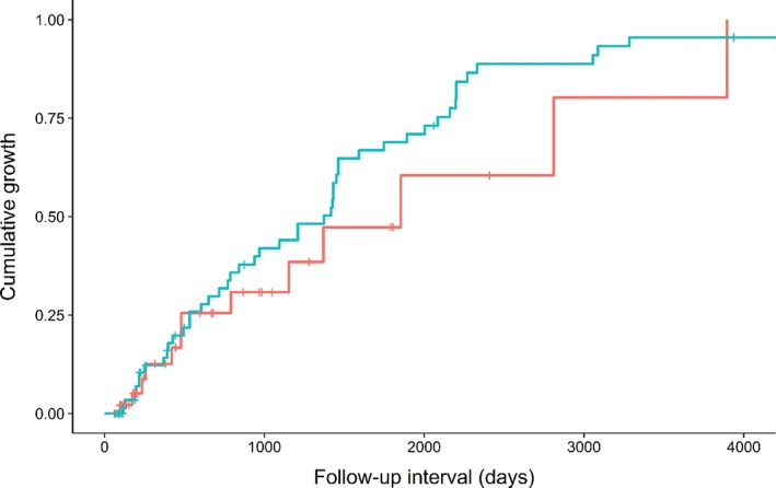
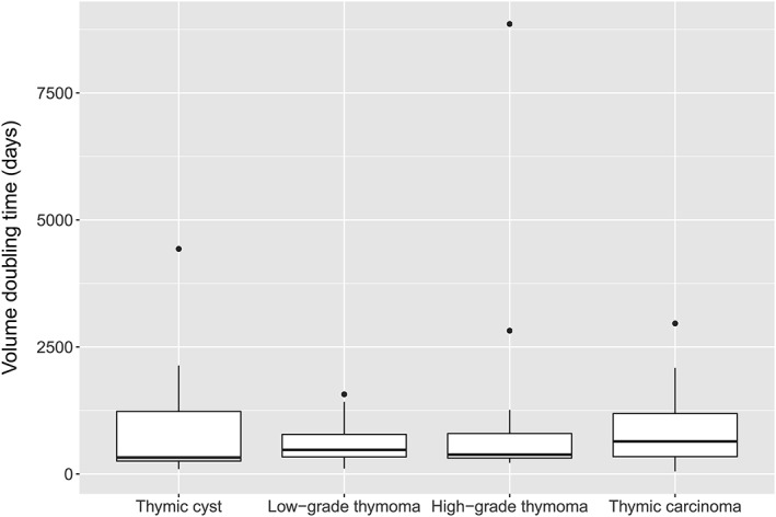
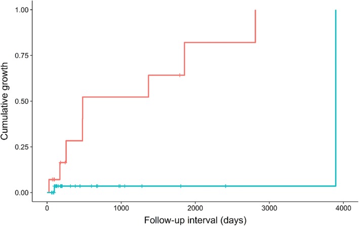
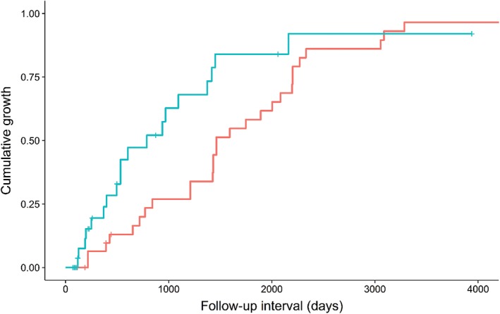
The frequency of growth did not differ significantly between TETs and thymic cysts (P = 0.279). The VDT of thymic cysts (median 324 days) was not significantly different from that of the TETs (median 475 days; P = 0.808). Water attenuation (≤ 20 Hounsfield units) predicted growth in thymic cysts (P = 0.016; hazard ratio 13.2, 95% confidence interval 1.6-107.3), while lesion size (> 17.2 mm) predicted growth in TETs (P = 0.008 for size, P = 0.029 for size*time). For the growing lesions, the positive and negative predictive values of water attenuation for thymic cysts were 93% and 80%, respectively.

CONCLUSION

The frequencies of interval growth and VDTs were indistinguishable between TETs and thymic cysts. Water attenuation and lesion size predicted growth in thymic cysts and TETs, respectively. Among the growing lesions, water attenuation was a differential feature of thymic cysts.

摘要

背景

研究了胸腺瘤(TETs)和胸囊的生长速度,以确定它们是否可以区分,并确定临床和影像学的间隔生长预测因子。

方法

本回顾性研究纳入了 122 例经病理证实的胸囊(n=56)或 TETs(n=66)患者,他们至少间隔 8 周进行了两次连续的胸部 CT 扫描。测量平均直径和衰减值,计算体积倍增时间(VDT),并记录临床特征。使用对数秩检验比较 VDT。使用对数秩检验和 Cox 回归分析分析生长的预测因子。

结果

TETs 和胸囊的生长频率无显著差异(P=0.279)。胸囊的 VDT(中位数 324 天)与 TETs 的 VDT(中位数 475 天;P=0.808)无显著差异。水衰减(≤20 亨氏单位)预测胸囊的生长(P=0.016;危险比 13.2,95%置信区间 1.6-107.3),而病变大小(>17.2mm)预测 TETs 的生长(P=0.008 用于大小,P=0.029 用于大小*时间)。对于生长的病变,水衰减对胸囊的阳性和阴性预测值分别为 93%和 80%。

结论

TETs 和胸囊的间隔生长频率和 VDT 无显著差异。水衰减和病变大小分别预测了胸囊和 TETs 的生长。在生长的病变中,水衰减是胸囊的一个鉴别特征。

https://cdn.ncbi.nlm.nih.gov/pmc/blobs/198f/6449257/ae2c178f74b3/TCA-10-864-g006.jpg
https://cdn.ncbi.nlm.nih.gov/pmc/blobs/198f/6449257/09c7ca62299b/TCA-10-864-g001.jpg
https://cdn.ncbi.nlm.nih.gov/pmc/blobs/198f/6449257/1a9e4e1dd511/TCA-10-864-g002.jpg
https://cdn.ncbi.nlm.nih.gov/pmc/blobs/198f/6449257/1fcf86c56dfe/TCA-10-864-g003.jpg
https://cdn.ncbi.nlm.nih.gov/pmc/blobs/198f/6449257/77cbc0e394ff/TCA-10-864-g004.jpg
https://cdn.ncbi.nlm.nih.gov/pmc/blobs/198f/6449257/700c80d65078/TCA-10-864-g005.jpg
https://cdn.ncbi.nlm.nih.gov/pmc/blobs/198f/6449257/ae2c178f74b3/TCA-10-864-g006.jpg

相似文献

1
Growth of thymic epithelial tumors and thymic cysts: Differential radiological points.胸腺上皮肿瘤和胸腺囊肿的生长:放射学差异要点。
Thorac Cancer. 2019 Apr;10(4):864-871. doi: 10.1111/1759-7714.13016. Epub 2019 Feb 21.
2
Computed tomography radiomic feature analysis of thymic epithelial tumors: Differentiation of thymic epithelial tumors from thymic cysts and prediction of histological subtypes.计算机断层扫描放射组学特征分析胸腺瘤:胸腺瘤与胸囊肿的鉴别及组织学分型预测。
Jpn J Radiol. 2024 Apr;42(4):367-373. doi: 10.1007/s11604-023-01512-0. Epub 2023 Nov 27.
3
Role of quantitative energy spectrum CT parameters in differentiating thymic epithelial tumours and thymic cysts.定量能谱 CT 参数在胸腺瘤和胸囊肿鉴别诊断中的作用。
Clin Radiol. 2022 Feb;77(2):136-141. doi: 10.1016/j.crad.2021.10.017. Epub 2021 Nov 29.
4
JOURNAL CLUB: Doubling Time of Thymic Epithelial Tumors Correlates With World Health Organization Histopathologic Classification.期刊俱乐部:胸腺瘤倍增时间与世界卫生组织组织病理学分类相关。
AJR Am J Roentgenol. 2017 Oct;209(4):W202-W210. doi: 10.2214/AJR.17.17836. Epub 2017 Aug 10.
5
Distinguishing between Thymic Epithelial Tumors and Benign Cysts via Computed Tomography.通过计算机断层扫描鉴别胸腺瘤和良性囊肿。
Korean J Radiol. 2019 Apr;20(4):671-682. doi: 10.3348/kjr.2018.0400.
6
Doubling time of thymic epithelial tumours on CT: correlation with histological subtype.胸腺上皮肿瘤的 CT 倍增时间:与组织学亚型的相关性。
Eur Radiol. 2017 Oct;27(10):4030-4036. doi: 10.1007/s00330-017-4795-y. Epub 2017 Mar 22.
7
Differential diagnosis of thymic epithelial neoplasms on computed tomography using the diameter of the thymic vein.基于胸腺静脉直径对胸腺癌的 CT 鉴别诊断
Medicine (Baltimore). 2021 Nov 19;100(46):e27942. doi: 10.1097/MD.0000000000027942.
8
Volume-based quantification using dual-energy computed tomography in the differentiation of thymic epithelial tumours: an initial experience.基于容积的双能计算机断层扫描在胸腺上皮肿瘤鉴别诊断中的应用:初步经验
Eur Radiol. 2017 May;27(5):1992-2001. doi: 10.1007/s00330-016-4542-9. Epub 2016 Aug 23.
9
A radiomics model to predict the invasiveness of thymic epithelial tumors based on contrast‑enhanced computed tomography.基于增强 CT 的影像组学模型预测胸腺瘤侵袭性
Oncol Rep. 2020 Apr;43(4):1256-1266. doi: 10.3892/or.2020.7497. Epub 2020 Feb 11.
10
A tour of the thymus: a review of thymic lesions with radiologic and pathologic correlation.胸腺探秘:胸腺病变的影像学与病理学相关性综述
Can Assoc Radiol J. 2015 Feb;66(1):5-15. doi: 10.1016/j.carj.2013.09.003. Epub 2014 Apr 13.

引用本文的文献

1
Differentiation and characterization of non-thymomatous ocular myasthenia gravis children with CT.非胸腺瘤性儿童眼肌型重症肌无力的CT鉴别与特征分析
Acta Neurol Belg. 2025 Feb;125(1):149-156. doi: 10.1007/s13760-024-02669-9. Epub 2024 Oct 30.
2
Robotic surgery for thymic cysts: clinical features, management, and results of a multicentric study.胸腺囊肿的机器人手术:多中心研究的临床特征、治疗及结果
Updates Surg. 2024 May 30. doi: 10.1007/s13304-024-01895-3.
3
The Rarest of Rare Thymic Lesions: A 10-Year Surgical Pathology Experience.

本文引用的文献

1
Methodologic Guide for Evaluating Clinical Performance and Effect of Artificial Intelligence Technology for Medical Diagnosis and Prediction.医学诊断和预测人工智能技术临床效能评估的方法学指南
Radiology. 2018 Mar;286(3):800-809. doi: 10.1148/radiol.2017171920. Epub 2018 Jan 8.
2
Incidental Anterior Mediastinal Nodular Lesions on Chest CT in Asymptomatic Subjects.无症状受检者胸部 CT 偶然发现前纵隔结节性病变
J Thorac Oncol. 2018 Mar;13(3):359-366. doi: 10.1016/j.jtho.2017.11.124. Epub 2017 Dec 9.
3
JOURNAL CLUB: Doubling Time of Thymic Epithelial Tumors Correlates With World Health Organization Histopathologic Classification.
最罕见的胸腺病变:十年手术病理经验
Cancers (Basel). 2021 Aug 12;13(16):4056. doi: 10.3390/cancers13164056.
4
Clinical Experience with Thymic Cystectomy: A Single-Institution Study of 117 Cases from 2013 to 2019.胸腺囊肿切除术的临床经验:2013 年至 2019 年单中心 117 例研究。
Med Sci Monit. 2020 May 20;26:e923967. doi: 10.12659/MSM.923967.
期刊俱乐部:胸腺瘤倍增时间与世界卫生组织组织病理学分类相关。
AJR Am J Roentgenol. 2017 Oct;209(4):W202-W210. doi: 10.2214/AJR.17.17836. Epub 2017 Aug 10.
4
Doubling time of thymic epithelial tumours on CT: correlation with histological subtype.胸腺上皮肿瘤的 CT 倍增时间:与组织学亚型的相关性。
Eur Radiol. 2017 Oct;27(10):4030-4036. doi: 10.1007/s00330-017-4795-y. Epub 2017 Mar 22.
5
The 2015 World Health Organization Classification of Tumors of the Thymus: Continuity and Changes.《2015年世界卫生组织胸腺肿瘤分类:延续性与变化》
J Thorac Oncol. 2015 Oct;10(10):1383-95. doi: 10.1097/JTO.0000000000000654.
6
Introduction to The 2015 World Health Organization Classification of Tumors of the Lung, Pleura, Thymus, and Heart.《2015年世界卫生组织肺、胸膜、胸腺和心脏肿瘤分类》简介
J Thorac Oncol. 2015 Sep;10(9):1240-1242. doi: 10.1097/JTO.0000000000000663.
7
MR Imaging of Mediastinal Masses.纵隔肿块的磁共振成像
Magn Reson Imaging Clin N Am. 2015 May;23(2):141-64. doi: 10.1016/j.mric.2015.01.002.
8
Value of Computerized 3D Shape Analysis in Differentiating Encapsulated from Invasive Thymomas.计算机三维形状分析在鉴别包膜型与浸润型胸腺瘤中的价值
PLoS One. 2015 May 4;10(5):e0126175. doi: 10.1371/journal.pone.0126175. eCollection 2015.
9
Anterior Mediastinal Masses in the Framingham Heart Study: Prevalence and CT Image Characteristics.弗雷明汉心脏研究中的前纵隔肿块:患病率及CT图像特征
Eur J Radiol Open. 2015;2:26-31. doi: 10.1016/j.ejro.2014.12.003.
10
The impact of thymoma histotype on prognosis in a worldwide database.全球数据库中胸腺瘤组织学类型对预后的影响。
J Thorac Oncol. 2015 Feb;10(2):367-72. doi: 10.1097/JTO.0000000000000393.