Araki Tetsuro, Nishino Mizuki, Gao Wei, Dupuis Josée, Washko George R, Hunninghake Gary M, Murakami Takamichi, O'Connor George T, Hatabu Hiroto
Department of Radiology, Center for Pulmonary Functional Imaging, Brigham and Women's Hospital, Harvard Medical School, Boston, MA ; Department of Radiology, Kinki University Faculty of Medicine, Osaka-Sayama, Japan.
Department of Radiology, Center for Pulmonary Functional Imaging, Brigham and Women's Hospital, Harvard Medical School, Boston, MA.
Eur J Radiol Open. 2015;2:26-31. doi: 10.1016/j.ejro.2014.12.003.
To investigate the prevalence and CT image characteristics of anterior mediastinal masses in a population-based cohort and their association with the demographics of the participants.
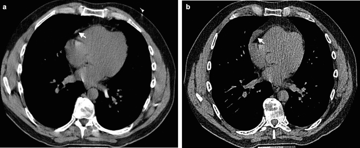
Chest CT scans of 2571 Framingham Heart Study participants (mean age 58.9 years, 51% female) were evaluated by two board-certified radiologists with expertise in thoracic imaging for the presence of anterior mediastinal masses, their shape, contour, location, invasion of adjacent structures, fat content, and calcification. For participants with anterior mediastinal masses, a previous cardiac CT scan was reviewed for interval size change of the masses, when available. The demographics of the participants were studied for any association with the presence of anterior mediastinal masses.
Of 2571, 23 participants (0.9%, 95% CI: 0.6 to 1.3) had anterior mediastinal masses on CT. The most common CT characteristics were oval shape, lobular contour, and midline location, showing soft tissue density (median 32.1 HU). Fat content was detected in a few cases (9%, 2/23). Six out of eight masses with available prior cardiac CT scans demonstrated an interval growth over a median period of 6.5 years. No risk factors for anterior mediastinal masses were detected among participants' demographics, including age, sex, BMI, and cigarette smoking.
The prevalence of anterior mediastinal masses is 0.9% in the Framingham Heart Study. Those masses may increase in size when observed over 5-7 years. Investigation of clinical significance in incidentally found anterior mediastinal masses with a longer period of follow-up would be necessary.
在一个基于人群的队列中调查前纵隔肿块的患病率、CT图像特征及其与参与者人口统计学特征的关联。
由两位具有胸部成像专业知识的认证放射科医生对2571名弗雷明汉心脏研究参与者(平均年龄58.9岁,51%为女性)的胸部CT扫描进行评估,以确定前纵隔肿块的存在、其形状、轮廓、位置、对相邻结构的侵犯、脂肪含量和钙化情况。对于患有前纵隔肿块的参与者,如有可用的先前心脏CT扫描,则对肿块的间隔大小变化进行回顾。研究参与者的人口统计学特征与前纵隔肿块存在之间的任何关联。
在2571名参与者中,23名(0.9%,95%CI:0.6至1.3)在CT上有前纵隔肿块。最常见的CT特征为椭圆形、分叶状轮廓和中线位置,表现为软组织密度(中位数32.1HU)。少数病例(9%,2/23)检测到脂肪含量。在八名有可用先前心脏CT扫描的肿块中,六名在中位期6.5年期间显示有间隔生长。在参与者的人口统计学特征中,包括年龄、性别、BMI和吸烟情况,未检测到前纵隔肿块的危险因素。
在弗雷明汉心脏研究中,前纵隔肿块的患病率为0.9%。这些肿块在5 - 7年的观察期内可能会增大。有必要对偶然发现的前纵隔肿块进行更长时间随访的临床意义进行研究。