• 文献检索
  • 文档翻译
  • 深度研究
  • 学术资讯
  • Suppr Zotero 插件Zotero 插件
  • 邀请有礼
  • 套餐&价格
  • 历史记录
应用&插件
Suppr Zotero 插件Zotero 插件浏览器插件Mac 客户端Windows 客户端微信小程序
定价
高级版会员购买积分包购买API积分包
服务
文献检索文档翻译深度研究API 文档MCP 服务
关于我们
关于 Suppr公司介绍联系我们用户协议隐私条款
关注我们

Suppr 超能文献

核心技术专利:CN118964589B侵权必究
粤ICP备2023148730 号-1Suppr @ 2026

文献检索

告别复杂PubMed语法,用中文像聊天一样搜索,搜遍4000万医学文献。AI智能推荐,让科研检索更轻松。

立即免费搜索

文件翻译

保留排版,准确专业,支持PDF/Word/PPT等文件格式,支持 12+语言互译。

免费翻译文档

深度研究

AI帮你快速写综述,25分钟生成高质量综述,智能提取关键信息,辅助科研写作。

立即免费体验

蛛网膜下腔出血后血脑屏障破坏的潜在治疗分子靶点。

Potential therapeutic molecular targets for blood-brain barrier disruption after subarachnoid hemorrhage.

作者信息

Kanamaru Hideki, Suzuki Hidenori

机构信息

Department of Neurosurgery, Mie University Graduate School of Medicine, Tsu, Japan.

出版信息

Neural Regen Res. 2019 Jul;14(7):1138-1143. doi: 10.4103/1673-5374.251190.

DOI:10.4103/1673-5374.251190
PMID:30804237
原文链接:https://pmc.ncbi.nlm.nih.gov/articles/PMC6425837/
Abstract

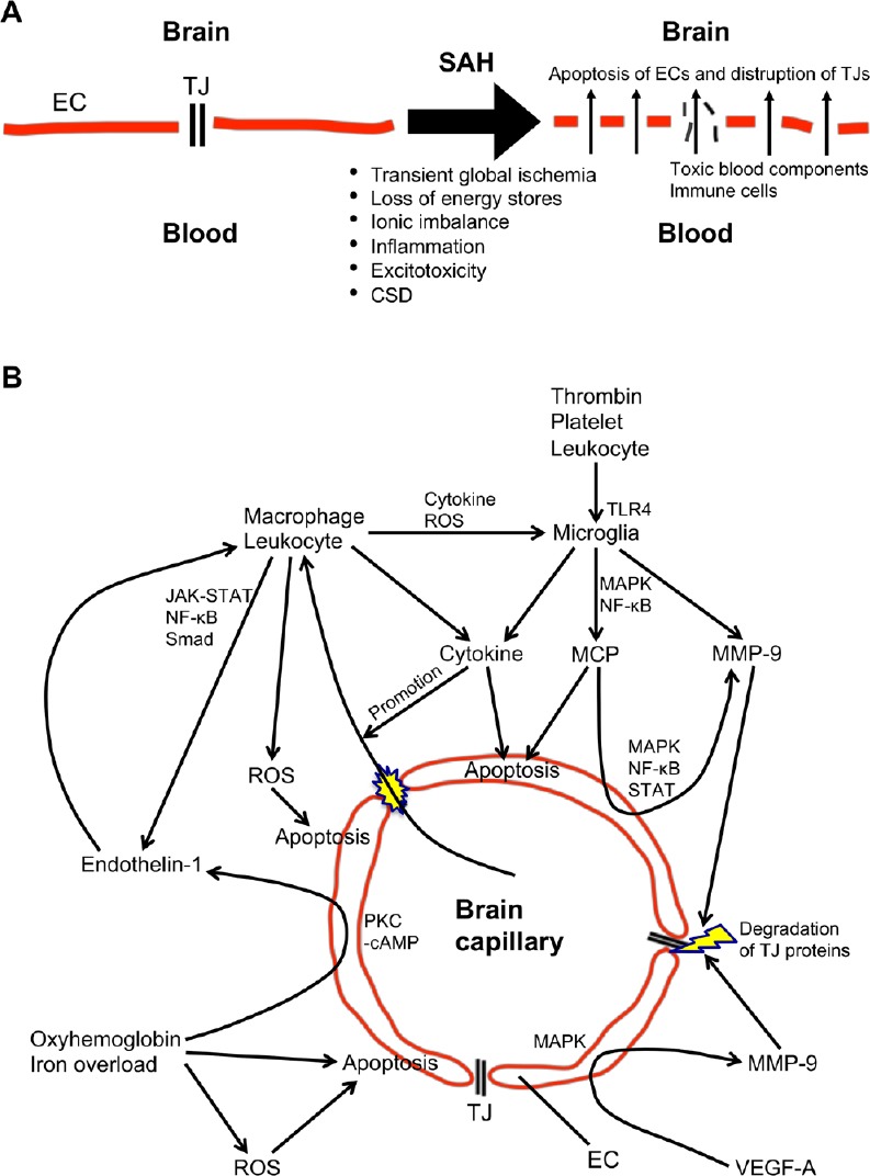
Aneurysmal subarachnoid hemorrhage remains serious hemorrhagic stroke with high morbidities and mortalities. Aneurysm rupture causes arterial bleeding-induced mechanical brain tissue injuries and elevated intracranial pressure, followed by global cerebral ischemia. Post-subarachnoid hemorrhage ischemia, tissue injuries as well as extravasated blood components and the breakdown products activate microglia, astrocytes and Toll-like receptor 4, and disrupt blood-brain barrier associated with the induction of many inflammatory and other cascades. Once blood-brain barrier is disrupted, brain tissues are directly exposed to harmful blood contents and immune cells, which aggravate brain injuries furthermore. Blood-brain barrier disruption after subarachnoid hemorrhage may be developed by a variety of mechanisms including endothelial cell apoptosis and disruption of tight junction proteins. Many molecules and pathways have been reported to disrupt the blood-brain barrier after subarachnoid hemorrhage, but the exact mechanisms remain unclear. Multiple independent and/or interconnected signaling pathways may be involved in blood-brain barrier disruption after subarachnoid hemorrhage. This review provides recent understandings of the mechanisms and the potential therapeutic targets of blood-brain barrier disruption after subarachnoid hemorrhage.

摘要

动脉瘤性蛛网膜下腔出血仍然是一种严重的出血性中风,具有很高的发病率和死亡率。动脉瘤破裂导致动脉出血引起的机械性脑组织损伤和颅内压升高,随后出现全脑缺血。蛛网膜下腔出血后的缺血、组织损伤以及外渗的血液成分和分解产物会激活小胶质细胞、星形胶质细胞和Toll样受体4,并破坏血脑屏障,引发许多炎症和其他级联反应。一旦血脑屏障被破坏,脑组织就会直接暴露于有害的血液成分和免疫细胞中,从而进一步加重脑损伤。蛛网膜下腔出血后血脑屏障的破坏可能是由多种机制引起的,包括内皮细胞凋亡和紧密连接蛋白的破坏。许多分子和信号通路已被报道与蛛网膜下腔出血后血脑屏障的破坏有关,但确切机制仍不清楚。多个独立和/或相互关联的信号通路可能参与蛛网膜下腔出血后血脑屏障的破坏。本文综述了蛛网膜下腔出血后血脑屏障破坏的机制及潜在治疗靶点的最新研究进展。

https://cdn.ncbi.nlm.nih.gov/pmc/blobs/c641/6425837/6d6f2742b232/NRR-14-1138-g001.jpg

相似文献

1
Potential therapeutic molecular targets for blood-brain barrier disruption after subarachnoid hemorrhage.蛛网膜下腔出血后血脑屏障破坏的潜在治疗分子靶点。
Neural Regen Res. 2019 Jul;14(7):1138-1143. doi: 10.4103/1673-5374.251190.
2
Underlying Mechanisms and Potential Therapeutic Molecular Targets in Blood-Brain Barrier Disruption after Subarachnoid Hemorrhage.蛛网膜下腔出血后血脑屏障破坏的潜在机制及治疗分子靶点
Curr Neuropharmacol. 2020;18(12):1168-1179. doi: 10.2174/1570159X18666200106154203.
3
Toll-Like Receptor 4 and Tenascin-C Signaling in Cerebral Vasospasm and Brain Injuries After Subarachnoid Hemorrhage.蛛网膜下腔出血后脑血管痉挛和脑损伤中的Toll样受体4和腱生蛋白-C信号传导
Acta Neurochir Suppl. 2020;127:91-96. doi: 10.1007/978-3-030-04615-6_15.
4
Toll-like receptor 4 as a possible therapeutic target for delayed brain injuries after aneurysmal subarachnoid hemorrhage.Toll样受体4作为动脉瘤性蛛网膜下腔出血后迟发性脑损伤的潜在治疗靶点。
Neural Regen Res. 2017 Feb;12(2):193-196. doi: 10.4103/1673-5374.200795.
5
Apigenin protects blood-brain barrier and ameliorates early brain injury by inhibiting TLR4-mediated inflammatory pathway in subarachnoid hemorrhage rats.芹菜素通过抑制蛛网膜下腔出血大鼠TLR4介导的炎症途径保护血脑屏障并改善早期脑损伤。
Int Immunopharmacol. 2015 Sep;28(1):79-87. doi: 10.1016/j.intimp.2015.05.024. Epub 2015 May 28.
6
Modified Citrus Pectin Prevents Blood-Brain Barrier Disruption in Mouse Subarachnoid Hemorrhage by Inhibiting Galectin-3.改性柑橘果胶通过抑制半乳糖凝集素-3防止小鼠蛛网膜下腔出血血脑屏障破坏。
Stroke. 2018 Nov;49(11):2743-2751. doi: 10.1161/STROKEAHA.118.021757.
7
Endothelial Cell Dysfunction and Injury in Subarachnoid Hemorrhage.蛛网膜下腔出血中的血管内皮细胞功能障碍与损伤。
Mol Neurobiol. 2019 Mar;56(3):1992-2006. doi: 10.1007/s12035-018-1213-7. Epub 2018 Jul 7.
8
The Role of Matricellular Proteins in Brain Edema after Subarachnoid Hemorrhage.基质细胞蛋白在蛛网膜下腔出血后脑水肿中的作用
Acta Neurochir Suppl. 2016;121:151-6. doi: 10.1007/978-3-319-18497-5_27.
9
Neuroinflammation and Microvascular Dysfunction After Experimental Subarachnoid Hemorrhage: Emerging Components of Early Brain Injury Related to Outcome.实验性蛛网膜下腔出血后的神经炎症和微血管功能障碍:与预后相关的早期脑损伤的新兴因素。
Neurocrit Care. 2019 Oct;31(2):373-389. doi: 10.1007/s12028-019-00710-x.
10
Inflammatory role of microglia in brain injury caused by subarachnoid hemorrhage.小胶质细胞在蛛网膜下腔出血所致脑损伤中的炎症作用
Front Cell Neurosci. 2022 Dec 6;16:956185. doi: 10.3389/fncel.2022.956185. eCollection 2022.

引用本文的文献

1
Tenascin-C as a Target for Intervention in Delayed Cerebral Ischemia After Subarachnoid Hemorrhage.腱生蛋白-C作为蛛网膜下腔出血后迟发性脑缺血干预靶点的研究
Acta Neurochir Suppl. 2025;136:11-17. doi: 10.1007/978-3-031-89844-0_2.
2
Acute Neurovascular Inflammatory Profile in Patients with Aneurysmal Subarachnoid Hemorrhage.动脉瘤性蛛网膜下腔出血患者的急性神经血管炎症特征
Biomolecules. 2025 Apr 23;15(5):613. doi: 10.3390/biom15050613.
3
Therapeutic potential of stem cells in subarachnoid hemorrhage.干细胞在蛛网膜下腔出血中的治疗潜力。

本文引用的文献

1
Lessons from tenascin-C knockout mice and potential clinical application to subarachnoid hemorrhage.来自腱生蛋白-C基因敲除小鼠的经验教训及在蛛网膜下腔出血中的潜在临床应用。
Neural Regen Res. 2019 Feb;14(2):262-264. doi: 10.4103/1673-5374.244789.
2
Modified Citrus Pectin Prevents Blood-Brain Barrier Disruption in Mouse Subarachnoid Hemorrhage by Inhibiting Galectin-3.改性柑橘果胶通过抑制半乳糖凝集素-3防止小鼠蛛网膜下腔出血血脑屏障破坏。
Stroke. 2018 Nov;49(11):2743-2751. doi: 10.1161/STROKEAHA.118.021757.
3
Tenascin-C in brain injuries and edema after subarachnoid hemorrhage: Findings from basic and clinical studies.
Neural Regen Res. 2025 Apr 1;20(4):936-945. doi: 10.4103/NRR.NRR-D-24-00124. Epub 2024 May 17.
4
Mitochondrial dysfunction and quality control lie at the heart of subarachnoid hemorrhage.线粒体功能障碍和质量控制是蛛网膜下腔出血的核心问题。
Neural Regen Res. 2024 Apr;19(4):825-832. doi: 10.4103/1673-5374.381493.
5
Inflammatory role of microglia in brain injury caused by subarachnoid hemorrhage.小胶质细胞在蛛网膜下腔出血所致脑损伤中的炎症作用
Front Cell Neurosci. 2022 Dec 6;16:956185. doi: 10.3389/fncel.2022.956185. eCollection 2022.
6
Progress in Research on TLR4-Mediated Inflammatory Response Mechanisms in Brain Injury after Subarachnoid Hemorrhage.蛛网膜下腔出血后脑损伤中 TLR4 介导的炎症反应机制的研究进展。
Cells. 2022 Nov 26;11(23):3781. doi: 10.3390/cells11233781.
7
The protective effect of low-dose minocycline on brain microvascular ultrastructure in a rodent model of subarachnoid hemorrhage.小剂量米诺环素对蛛网膜下腔出血啮齿动物模型脑微血管超微结构的保护作用。
Histochem Cell Biol. 2023 Jan;159(1):91-114. doi: 10.1007/s00418-022-02150-9. Epub 2022 Sep 24.
8
Roles of glutamate in brain injuries after subarachnoid hemorrhage.谷氨酸在蛛网膜下腔出血后脑损伤中的作用。
Histol Histopathol. 2022 Nov;37(11):1041-1051. doi: 10.14670/HH-18-509. Epub 2022 Sep 6.
9
Selective sphingosine-1-phosphate receptor 1 modulator attenuates blood-brain barrier disruption following traumatic brain injury by inhibiting vesicular transcytosis.选择性鞘氨醇-1-磷酸受体 1 调节剂通过抑制囊泡转胞吞作用来减轻创伤性脑损伤后的血脑屏障破坏。
Fluids Barriers CNS. 2022 Jul 11;19(1):57. doi: 10.1186/s12987-022-00356-6.
10
Melatonin as a Potential Neuroprotectant: Mechanisms in Subarachnoid Hemorrhage-Induced Early Brain Injury.褪黑素作为一种潜在的神经保护剂:蛛网膜下腔出血所致早期脑损伤的机制
Front Aging Neurosci. 2022 Apr 29;14:899678. doi: 10.3389/fnagi.2022.899678. eCollection 2022.
蛛网膜下腔出血后脑损伤和水肿中的 tenascin-C:基础和临床研究的结果。
J Neurosci Res. 2020 Jan;98(1):42-56. doi: 10.1002/jnr.24330. Epub 2018 Sep 22.
4
Systematic Review of In Vivo Animal Models of Subarachnoid Hemorrhage: Species, Standard Parameters, and Outcomes.蛛网膜下腔出血体内动物模型的系统评价:物种、标准参数及结果
Transl Stroke Res. 2018 Sep 12. doi: 10.1007/s12975-018-0657-4.
5
Blood-Brain Barrier Permeability in Aneurysmal Subarachnoid Hemorrhage: Correlation With Clinical Outcomes.颅内动脉瘤性蛛网膜下腔出血的血脑屏障通透性:与临床结局的相关性。
AJR Am J Roentgenol. 2018 Oct;211(4):891-895. doi: 10.2214/AJR.17.18237. Epub 2018 Aug 7.
6
Dose-Dependent Inhibitory Effects of Cilostazol on Delayed Cerebral Infarction After Aneurysmal Subarachnoid Hemorrhage.西洛他唑对动脉瘤性蛛网膜下腔出血后迟发性脑梗死的剂量依赖性抑制作用。
Transl Stroke Res. 2019 Aug;10(4):381-388. doi: 10.1007/s12975-018-0650-y. Epub 2018 Jul 23.
7
Matricellular proteins as possible biomarkers for early brain injury after aneurysmal subarachnoid hemorrhage.基质细胞蛋白作为动脉瘤性蛛网膜下腔出血后早期脑损伤的潜在生物标志物。
Neural Regen Res. 2018 Jul;13(7):1175-1178. doi: 10.4103/1673-5374.235022.
8
Endothelial Cell Dysfunction and Injury in Subarachnoid Hemorrhage.蛛网膜下腔出血中的血管内皮细胞功能障碍与损伤。
Mol Neurobiol. 2019 Mar;56(3):1992-2006. doi: 10.1007/s12035-018-1213-7. Epub 2018 Jul 7.
9
Selective Toll-Like Receptor 4 Antagonists Prevent Acute Blood-Brain Barrier Disruption After Subarachnoid Hemorrhage in Mice.选择性 Toll 样受体 4 拮抗剂可预防小鼠蛛网膜下腔出血后急性血脑屏障破坏。
Mol Neurobiol. 2019 Feb;56(2):976-985. doi: 10.1007/s12035-018-1145-2. Epub 2018 May 31.
10
Possible Role of Inflammation and Galectin-3 in Brain Injury after Subarachnoid Hemorrhage.炎症和半乳糖凝集素-3在蛛网膜下腔出血后脑损伤中的可能作用
Brain Sci. 2018 Feb 7;8(2):30. doi: 10.3390/brainsci8020030.