Pescatori Lorenzo Carlo, Savarino Edoardo, Mauri Giovanni, Silvestri Enzo, Cariati Maurizio, Sardanelli Francesco, Sconfienza Luca Maria
Università degli Studi di Milano, Milano, Italia.
Università di Padova, Padova, Italia.
Radiol Bras. 2019 Jan-Feb;52(1):1-6. doi: 10.1590/0100-3984.2017.0211.
To evaluate the feasibility of quantifying visceral adipose tissue (VAT) on computed tomography (CT) and magnetic resonance imaging (MRI) scans, using freeware, as well as calculating intraobserver and interobserver reproducibility.
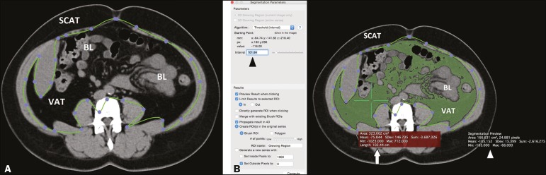
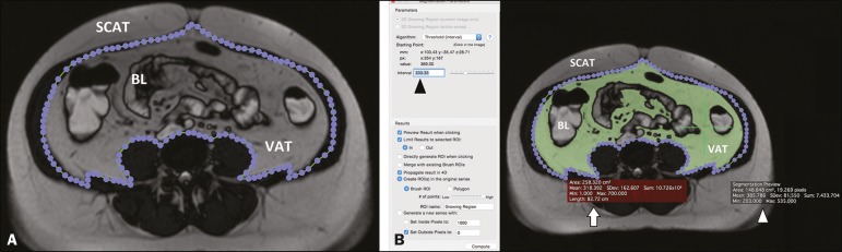
We quantified VAT in patients who underwent abdominal CT and MRI at our institution between 2010 and 2015, with a maximum of three months between the two examinations. A slice acquired at the level of the umbilicus was selected. Segmentation was performed with the region growing algorithm of the freeware employed. Intraobserver and interobserver reproducibility were evaluated, as was the accuracy of MRI in relation to that of CT.
Thirty-one patients (14 males and 17 females; mean age of 57 ± 15 years) underwent CT and MRI (mean interval between the examinations, 28 ± 12 days). The interobserver reproducibility was 82% for CT (bias = 1.52 cm; = 0.488), 86% for T1-weighted MRI (bias = -4.36 cm; = 0.006), and 88% for T2-weighted MRI (bias = -0.52 cm; = 0.735). The intraobserver reproducibility was 90% for CT (bias = 0.14 cm; = 0.912), 92% for T1-weighted MRI (bias = -3,4 cm; = 0.035), and 90% for T2-weighted MRI (bias = -0.30 cm; = 0.887). The reproducibility between T1-weighted MRI and T2-weighted MRI was 87% (bias = -0.11 cm; = 0.957). In comparison with the accuracy of CT, that of T1-weighted and T2-weighted MRI was 89% and 91%, respectively.
The program employed can be used in order to quantify VAT on CT, T1-weighted MRI, and T2-weighted MRI scans. Overall, the accuracy of MRI (in comparison with that of CT) appears to be high, as do intraobserver and interobserver reproducibility. However, the quantification of VAT seems to be less reproducible in T1-weighted sequences.
评估使用免费软件在计算机断层扫描(CT)和磁共振成像(MRI)扫描上对内脏脂肪组织(VAT)进行量化的可行性,以及计算观察者内和观察者间的可重复性。
我们对2010年至2015年间在本机构接受腹部CT和MRI检查的患者的VAT进行了量化,两次检查之间的间隔最长为三个月。选择在脐水平获取的切片。使用所采用免费软件的区域生长算法进行分割。评估了观察者内和观察者间的可重复性,以及MRI相对于CT的准确性。
31例患者(14例男性和17例女性;平均年龄57±15岁)接受了CT和MRI检查(检查之间的平均间隔为28±12天)。CT的观察者间可重复性为82%(偏差 = 1.52 cm; = 0.488),T1加权MRI为86%(偏差 = -4.36 cm; = 0.006),T2加权MRI为88%(偏差 = -0.52 cm; = 0.735)。CT的观察者内可重复性为90%(偏差 = 0.14 cm; = 0.912),T1加权MRI为92%(偏差 = -3.4 cm; = 0.035),T2加权MRI为90%(偏差 = -0.30 cm; = 0.887)。T1加权MRI和T2加权MRI之间的可重复性为87%(偏差 = -0.11 cm; = 0.957)。与CT的准确性相比,T1加权和T2加权MRI的准确性分别为89%和91%。
所采用的程序可用于在CT、T1加权MRI和T2加权MRI扫描上对VAT进行量化。总体而言,MRI(与CT相比)的准确性似乎较高,观察者内和观察者间的可重复性也是如此。然而,VAT的量化在T1加权序列中似乎重复性较差。