Bereiter David A, Thompson Randall, Rahman Mostafeezur
Department of Diagnostic and Biological Sciences, University of Minnesota School of Dentistry, Minneapolis, MN, United States.
Front Integr Neurosci. 2019 Feb 12;13:3. doi: 10.3389/fnint.2019.00003. eCollection 2019.
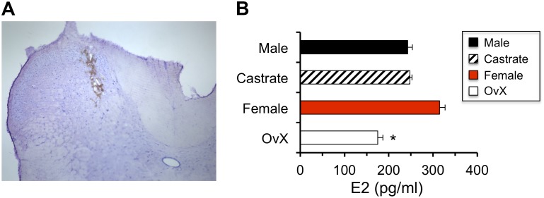
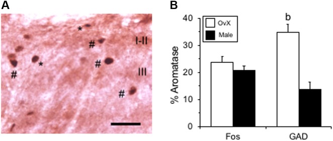
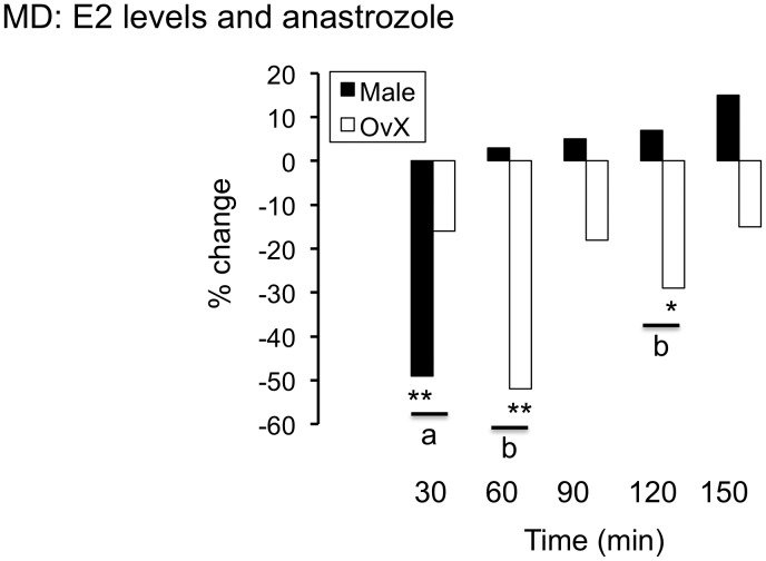
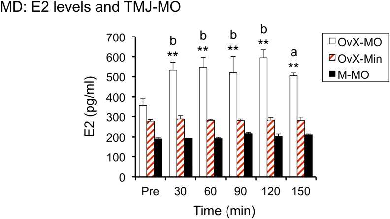
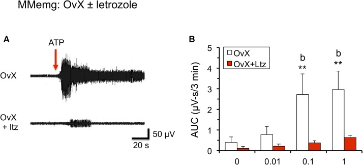
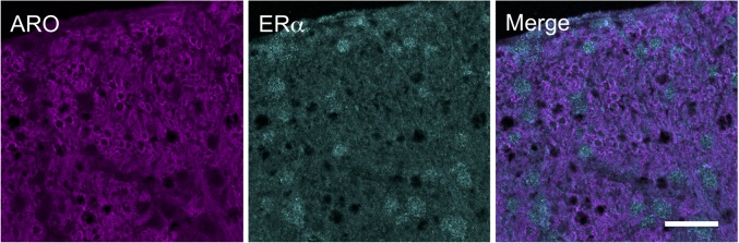
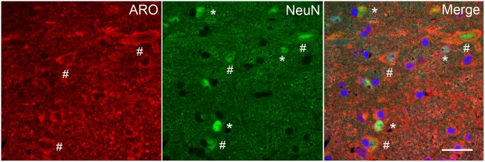
Estrogen status is a significant risk factor in the development of temporomandibular joint disorders (TMD). Classically, estrogen status is thought to derive mainly from ovarian sources; however, it is well known that estradiol (E2) also is synthesized by neurons in the brain. This study tested the hypothesis that E2 is produced by neurons in trigeminal subnucleus caudalis (Vc), the principal site of termination for sensory afferents that supply the temporomandibular joint (TMJ), to modify evoked responses in a model of TMJ nociception in male and female rats. Intra-TMJ injection of the small fiber excitant, allyl isothiocyanate (AIC), increased the levels of E2 collected from microdialysis probes sites at Vc of ovariectomized (OvX) female rats, ipsilateral to the stimulus, whereas males displayed no change. Dialysate levels of E2 collected from probe sites in the contralateral Vc or cerebellum in OvX rats were not affected by TMJ stimulation. Reverse dialysis of anastrozole, an aromatase (ARO) inhibitor, via the probe reduced perfusate levels of E2 in Vc. Systemic administration of letrozole, a non-steroid ARO inhibitor, for 4 days prevented TMJ-evoked increases in masseter muscle electromyography (MMemg) activity. ARO-positive neurons were distributed mainly in superficial laminae (I-III) at Vc and cell counts revealed no significant difference between OvX and male rats. Intra-TMJ injection of AIC revealed similar numbers of ARO/Fos dual-labeled neurons in OvX and male rats. By contrast, the percentage of ARO neurons co-labeled for glutamic acid decarboxylase (GAD), the biosynthetic enzyme for GABA, was greater in OvX (35%) than male rats (14%). Few ARO-positive neurons were co-labeled for estrogen receptor alpha. These data indicate that E2 is secreted continuously by Vc neurons and that acute stimulation of TMJ nociceptors evokes further secretion in a sex-dependent manner. Reduced TMJ-evoked MMemg activity after ARO inhibition suggests that locally produced E2 by Vc neurons acts via paracrine mechanisms to modify TMJ nociception in female rats.
雌激素状态是颞下颌关节紊乱病(TMD)发生发展的一个重要风险因素。传统上,雌激素状态被认为主要来源于卵巢;然而,众所周知,雌二醇(E2)也由大脑中的神经元合成。本研究检验了以下假设:E2由三叉神经尾侧亚核(Vc)中的神经元产生,Vc是供应颞下颌关节(TMJ)的感觉传入纤维的主要终止部位,它可改变雄性和雌性大鼠TMJ伤害感受模型中的诱发反应。向TMJ内注射小纤维刺激剂异硫氰酸烯丙酯(AIC),可使去卵巢(OvX)雌性大鼠刺激同侧Vc处微透析探针位点收集的E2水平升高,而雄性大鼠则无变化。OvX大鼠对侧Vc或小脑中探针位点收集的E2透析液水平不受TMJ刺激的影响。通过探针反向透析芳香化酶(ARO)抑制剂阿那曲唑可降低Vc中E2的灌流液水平。连续4天全身给予非甾体ARO抑制剂来曲唑可防止TMJ诱发的咬肌肌电图(MMemg)活动增加。ARO阳性神经元主要分布在Vc的浅层(I - III层),细胞计数显示OvX大鼠和雄性大鼠之间无显著差异。向TMJ内注射AIC显示,OvX大鼠和雄性大鼠中ARO/Fos双标神经元数量相似。相比之下,与γ-氨基丁酸(GABA)生物合成酶谷氨酸脱羧酶(GAD)共标记的ARO神经元百分比,OvX大鼠(35%)高于雄性大鼠(14%)。很少有ARO阳性神经元与雌激素受体α共标记。这些数据表明,E2由Vc神经元持续分泌,TMJ伤害感受器的急性刺激以性别依赖的方式引起进一步分泌。ARO抑制后TMJ诱发的MMemg活动降低表明,Vc神经元局部产生的E2通过旁分泌机制作用于雌性大鼠的TMJ伤害感受。