Koelsche Christian, Stichel Damian, Griewank Klaus G, Schrimpf Daniel, Reuss David E, Bewerunge-Hudler Melanie, Vokuhl Christian, Dinjens Winand N M, Petersen Iver, Mittelbronn Michel, Cuevas-Bourdier Adrian, Buslei Rolf, Pfister Stefan M, Flucke Uta, Mechtersheimer Gunhild, Mentzel Thomas, von Deimling Andreas
1Department of General Pathology, Institute of Pathology, Heidelberg University Hospital, Im Neuenheimer Feld 224, 69120 Heidelberg, Baden-Württemberg Germany.
2Department of Neuropathology, Institute of Pathology, Heidelberg University Hospital, Im Neuenheimer Feld 224, 69120 Heidelberg, Baden-Württemberg Germany.
Clin Sarcoma Res. 2019 Feb 14;9:2. doi: 10.1186/s13569-019-0113-6. eCollection 2019.
Atypical fibroxanthomas (AFX) and pleomorphic dermal sarcomas (PDS) are lesions of the skin with overlapping histologic features and unspecific molecular traits. PDS behaves aggressive compared to AFX. Thus, a precise delineation, although challenging in some instances, is relevant.
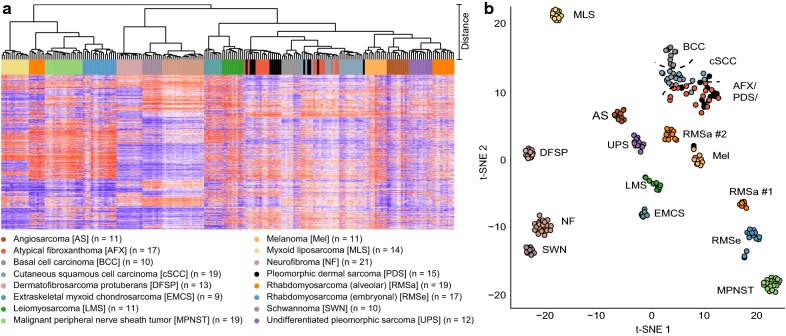
We examined the value of DNA-methylation profiling and copy number analysis for separating these tumors. DNA-methylation data were generated from 17 AFX and 15 PDS using the Illumina EPIC array. These were compared with DNA-methylation data generated from 196 tumors encompassing potential histologic mimics like cutaneous squamous carcinomas (cSCC; n = 19), basal cell carcinomas (n = 10), melanoma metastases originating from the skin (n = 11), leiomyosarcomas (n = 11), angiosarcomas of the skin and soft tissue (n = 11), malignant peripheral nerve sheath tumors (n = 19), dermatofibrosarcomas protuberans (n = 13), extraskeletal myxoid chondrosarcomas (n = 9), myxoid liposarcomas (n = 14), schwannomas (n = 10), neurofibromas (n = 21), alveolar (n = 19) and embryonal (n = 17) rhabdomyosarcomas as well as undifferentiated pleomorphic sarcomas (n = 12).
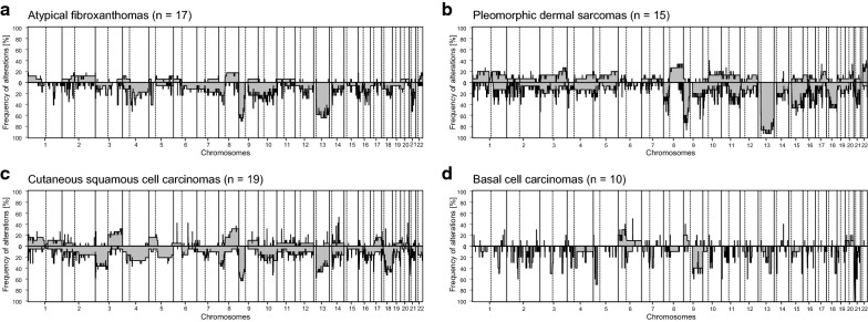
DNA-methylation profiling did not separate AFX from PDS. The DNA-methylation profiles of the other cases, however, were distinct from AFX/PDS. They reliably assigned to subtype-specific DNA-methylation clusters, although overlap occurred between some AFX/PDS and cSCC. Copy number profiling revealed alterations in a similar frequency and distribution between AFX and PDS. They involved losses of 9p (22/32) and 13q (25/32). Gains frequently involved 8q (8/32). Notably, a homozygous deletion of was more frequent in PDS (6/15) than in AFX (2/17), whereas amplifications were non-recurrent and overall rare (5/32).
Our findings support the concept that AFX and PDS belong to a common tumor spectrum. We could demonstrate the diagnostic value of DNA-methylation profiling to delineating AFX/PDS from potential mimics. However, the assessment of certain histologic features remains crucial for separating PDS from AFX.
非典型纤维黄色瘤(AFX)和多形性皮肤肉瘤(PDS)是具有重叠组织学特征和非特异性分子特征的皮肤病变。与AFX相比,PDS具有侵袭性。因此,尽管在某些情况下具有挑战性,但精确区分仍具有重要意义。
我们研究了DNA甲基化谱分析和拷贝数分析在区分这些肿瘤中的价值。使用Illumina EPIC阵列从17例AFX和15例PDS中生成DNA甲基化数据。将这些数据与从196例肿瘤中生成的DNA甲基化数据进行比较,这些肿瘤包括潜在的组织学相似病变,如皮肤鳞状细胞癌(cSCC;n = 19)、基底细胞癌(n = 10)、源自皮肤的黑色素瘤转移灶(n = 11)、平滑肌肉瘤(n = 11)、皮肤和软组织血管肉瘤(n = 11)、恶性外周神经鞘瘤(n = 19)、隆突性皮肤纤维肉瘤(n = 13)、骨外黏液样软骨肉瘤(n = 9)、黏液样脂肪肉瘤(n = 14)、神经鞘瘤(n = 10)、神经纤维瘤(n = 21)、肺泡型(n = 19)和胚胎型(n = 17)横纹肌肉瘤以及未分化多形性肉瘤(n = 12)。
DNA甲基化谱分析未能将AFX与PDS区分开来。然而,其他病例的DNA甲基化谱与AFX/PDS不同。尽管某些AFX/PDS与cSCC之间存在重叠,但它们可靠地归入亚型特异性DNA甲基化簇。拷贝数谱分析显示AFX和PDS之间的改变频率和分布相似。它们涉及9p(22/32)和13q(25/32)的缺失。增益常见于8q(8/32)。值得注意的是,PDS(6/15)中纯合缺失的频率高于AFX(2/17),而扩增不常见且总体罕见(5/32)。
我们的研究结果支持AFX和PDS属于共同肿瘤谱系的概念。我们能够证明DNA甲基化谱分析在将AFX/PDS与潜在相似病变区分开来方面的诊断价值。然而,评估某些组织学特征对于将PDS与AFX区分开来仍然至关重要。