• 文献检索
  • 文档翻译
  • 深度研究
  • 学术资讯
  • Suppr Zotero 插件Zotero 插件
  • 邀请有礼
  • 套餐&价格
  • 历史记录
应用&插件
Suppr Zotero 插件Zotero 插件浏览器插件Mac 客户端Windows 客户端微信小程序
定价
高级版会员购买积分包购买API积分包
服务
文献检索文档翻译深度研究API 文档MCP 服务
关于我们
关于 Suppr公司介绍联系我们用户协议隐私条款
关注我们

Suppr 超能文献

核心技术专利:CN118964589B侵权必究
粤ICP备2023148730 号-1Suppr @ 2026

文献检索

告别复杂PubMed语法,用中文像聊天一样搜索,搜遍4000万医学文献。AI智能推荐,让科研检索更轻松。

立即免费搜索

文件翻译

保留排版,准确专业,支持PDF/Word/PPT等文件格式,支持 12+语言互译。

免费翻译文档

深度研究

AI帮你快速写综述,25分钟生成高质量综述,智能提取关键信息,辅助科研写作。

立即免费体验

多模态超声成像在鉴别 TI-RADS 4 类良恶性结节中的诊断价值。

Diagnostic value of multimodal ultrasound imaging in differentiating benign and malignant TI-RADS category 4 nodules.

机构信息

The Second School of Clinical Medicine, Southern Medical University, Guangzhou, Guangdong, People's Republic of China.

Department of Ultrasound, Guangdong Academy of Medical Sciences/Guangdong General Hospital, Guangzhou, Guangdong, People's Republic of China.

出版信息

Int J Clin Oncol. 2019 Jun;24(6):632-639. doi: 10.1007/s10147-019-01397-y. Epub 2019 Mar 1.

DOI:10.1007/s10147-019-01397-y
PMID:30825007
原文链接:https://pmc.ncbi.nlm.nih.gov/articles/PMC6525125/
Abstract

BACKGROUND

Differential diagnosis of benign and malignant thyroid imaging reporting and data system category 4 (TI-RADS-4) nodules can be difficult using conventional ultrasound (US). This study aimed to evaluate whether multimodal ultrasound imaging can improve differentiation and characterization of benign and malignant TI-RADS-4 nodules.

METHODS

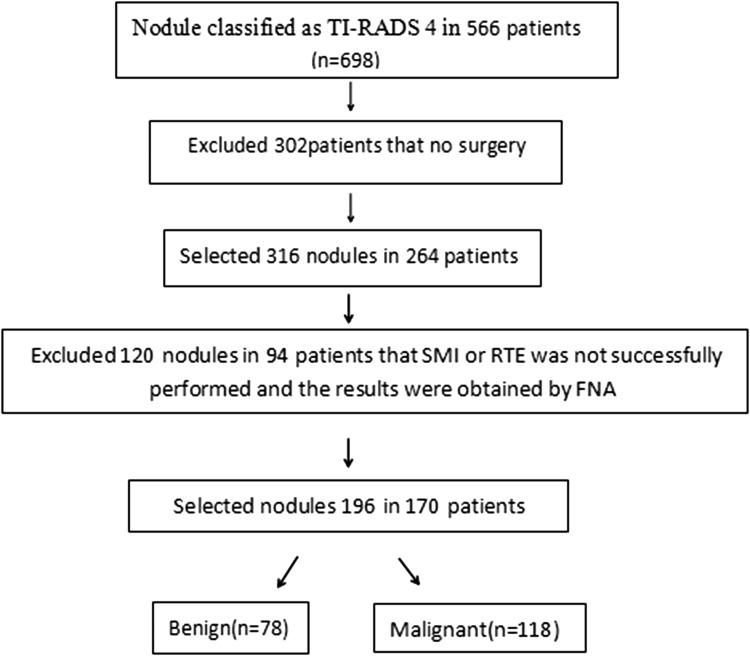
Multimodal ultrasound imaging, including US, superb microvascular imaging (SMI), and real-time elastography (RTE), were performed on 196 TI-RADS-4 nodules (78, benign; 118, malignant) in 170 consecutive patients. The sensitivity, specificity, accuracy, false negative rate (FNR), and false positive rate (FPR) of each single method and that of multimodal US imaging were determined by comparison with surgical pathology results.

RESULTS

The sensitivity, specificity, accuracy, FNR, and FPR for US were 65.25%, 69.23%, 66.84%, 34.75%, 30.77%, respectively; for SMI were 77.97%, 93.59%, 84.18%, 22.03%, 6.41%, respectively; RTE, 80.51%, 84.62%, 82.14%, 19.49%, 15.38%; and for multimodal US imaging were 94.08%, 87.18%, 91.33%, 6.93%, 12.82%, respectively. The areas under the received operating characteristic curve for US, SMI, RTE, and multimodal US imaging in evaluating benign and malignant TI-RADS-4 nodules were 67.2%, 84.40%, 86.60%, and 95.50%, respectively.

CONCLUSIONS

The initial clinical results suggest that multimodal US imaging improves the diagnostic accuracy of TI-RADS-4 nodules and provides additional information for differentiating malignant and benign nodules.

摘要

背景

使用传统超声(US)对甲状腺影像报告和数据系统分类 4 级(TI-RADS-4)结节的良恶性进行鉴别诊断可能较为困难。本研究旨在评估多模态超声成像是否能改善 TI-RADS-4 级良恶性结节的鉴别和特征分析。

方法

对 170 例连续患者的 196 个 TI-RADS-4 级结节(78 个良性,118 个恶性)进行多模态超声成像检查,包括 US、超微血流成像(SMI)和实时弹性成像(RTE)。将手术病理结果作为参照,确定每种单模态方法及多模态 US 成像的敏感性、特异性、准确性、假阴性率(FNR)和假阳性率(FPR)。

结果

US 的敏感性、特异性、准确性、FNR 和 FPR 分别为 65.25%、69.23%、66.84%、34.75%、30.77%;SMI 分别为 77.97%、93.59%、84.18%、22.03%、6.41%;RTE 分别为 80.51%、84.62%、82.14%、19.49%、15.38%;多模态 US 成像分别为 94.08%、87.18%、91.33%、6.93%、12.82%。US、SMI、RTE 和多模态 US 成像在评估 TI-RADS-4 级良恶性结节的受试者工作特征曲线下面积分别为 67.2%、84.40%、86.60%和 95.50%。

结论

初步临床结果表明,多模态 US 成像可提高 TI-RADS-4 级结节的诊断准确性,为良恶性结节的鉴别提供更多信息。

https://cdn.ncbi.nlm.nih.gov/pmc/blobs/689b/6525125/94a2cf923ff8/10147_2019_1397_Fig3_HTML.jpg
https://cdn.ncbi.nlm.nih.gov/pmc/blobs/689b/6525125/59a92906597a/10147_2019_1397_Fig1_HTML.jpg
https://cdn.ncbi.nlm.nih.gov/pmc/blobs/689b/6525125/94a2cf923ff8/10147_2019_1397_Fig3_HTML.jpg
https://cdn.ncbi.nlm.nih.gov/pmc/blobs/689b/6525125/59a92906597a/10147_2019_1397_Fig1_HTML.jpg
https://cdn.ncbi.nlm.nih.gov/pmc/blobs/689b/6525125/94a2cf923ff8/10147_2019_1397_Fig3_HTML.jpg

相似文献

1
Diagnostic value of multimodal ultrasound imaging in differentiating benign and malignant TI-RADS category 4 nodules.多模态超声成像在鉴别 TI-RADS 4 类良恶性结节中的诊断价值。
Int J Clin Oncol. 2019 Jun;24(6):632-639. doi: 10.1007/s10147-019-01397-y. Epub 2019 Mar 1.
2
The diagnostic value of combination of TI-RADS and ultrasound elastography in the differentiation of benign and malignant thyroid nodules.TI-RADS与超声弹性成像联合应用在鉴别甲状腺良恶性结节中的诊断价值。
Clin Imaging. 2016 Sep-Oct;40(5):913-6. doi: 10.1016/j.clinimag.2016.04.014. Epub 2016 Apr 30.
3
Incremental diagnostic value of shear wave elastography combined with contrast-enhanced ultrasound in TI-RADS category 4a and 4b nodules.剪切波弹性成像联合超声造影在TI-RADS 4a和4b类结节中的增量诊断价值
J Med Ultrason (2001). 2020 Jul;47(3):453-462. doi: 10.1007/s10396-020-01016-8. Epub 2020 Apr 18.
4
Combination of ultrasound elastography with TI-RADS in the diagnosis of small thyroid nodules (≤10 mm): A new method to increase the diagnostic performance.超声弹性成像联合 TI-RADS 在诊断直径≤10mm 的甲状腺小结节中的应用:一种提高诊断效能的新方法。
Eur J Radiol. 2018 Dec;109:33-40. doi: 10.1016/j.ejrad.2018.10.024. Epub 2018 Oct 28.
5
Additional Value of Superb Microvascular Imaging for Thyroid Nodule Classification with the Thyroid Imaging Reporting and Data System.超声造影在甲状腺影像报告和数据系统分类甲状腺结节中的附加价值。
Ultrasound Med Biol. 2019 Aug;45(8):2040-2048. doi: 10.1016/j.ultrasmedbio.2019.05.001. Epub 2019 May 24.
6
The value of TI-RADS combined with superb micro-vascular imagine in distinguishing benign and malignant thyroid nodules: A meta-analysis.TI-RADS 联合超声微血管成像对鉴别甲状腺良恶性结节的价值:一项荟萃分析。
PLoS One. 2022 Jan 18;17(1):e0261521. doi: 10.1371/journal.pone.0261521. eCollection 2022.
7
Clinical Study of the Prediction of Malignancy in Thyroid Nodules: Modified Score versus 2017 American College of Radiology's Thyroid Imaging Reporting and Data System Ultrasound Lexicon.甲状腺结节良恶性预测的临床研究:改良评分与 2017 年美国放射学会甲状腺影像报告和数据系统超声词汇的比较。
Ultrasound Med Biol. 2019 Jul;45(7):1627-1637. doi: 10.1016/j.ultrasmedbio.2019.03.014. Epub 2019 May 4.
8
Assessment of Virtual Touch Tissue Imaging Quantification and the Ultrasound Thyroid Imaging Reporting and Data System in Patients With Thyroid Nodules Referred for Biopsy.对因活检而转诊的甲状腺结节患者进行虚拟触诊组织成像定量分析及超声甲状腺影像报告和数据系统评估。
J Ultrasound Med. 2018 Mar;37(3):725-736. doi: 10.1002/jum.14413. Epub 2017 Sep 28.
9
Thyroid Imaging Reporting and Data System (TI-RADS): the impact of Quantitative Strain Elastography for better stratification of cancer risks.甲状腺影像报告和数据系统(TI-RADS):定量应变弹性成像对更好地分层癌症风险的影响。
Med Ultrason. 2015 Sep;17(3):327-32. doi: 10.11152/mu.2013.2066.173.dst.
10
Suggested amendment of TI-RADS classification of thyroid nodules by shear wave elastography.基于剪切波弹性成像对甲状腺影像报告和数据系统(TI-RADS)甲状腺结节分类的建议修正
Acta Radiol. 2020 Aug;61(8):1026-1033. doi: 10.1177/0284185119889567. Epub 2019 Dec 11.

引用本文的文献

1
Multiparametric ultrasound techniques are superior to AI-assisted ultrasound for assessment of solid thyroid nodules: a prospective study.多参数超声技术在评估实性甲状腺结节方面优于人工智能辅助超声:一项前瞻性研究。
Endocrine. 2025 Jul 10. doi: 10.1007/s12020-025-04306-8.
2
Enhancing diagnostic accuracy of American College of Radiology TI-RADS 4 nodules: nomogram models based on MRI morphological features.提高美国放射学会TI-RADS 4类结节的诊断准确性:基于MRI形态学特征的列线图模型
Quant Imaging Med Surg. 2025 Feb 1;15(2):1679-1693. doi: 10.21037/qims-24-1427. Epub 2025 Jan 20.
3
Diagnostic value of greyscale ultrasound combined with superb microvascular imaging in thyroid nodules: a systematic review and meta-analysis.

本文引用的文献

1
Combination of ultrasound elastography with TI-RADS in the diagnosis of small thyroid nodules (≤10 mm): A new method to increase the diagnostic performance.超声弹性成像联合 TI-RADS 在诊断直径≤10mm 的甲状腺小结节中的应用:一种提高诊断效能的新方法。
Eur J Radiol. 2018 Dec;109:33-40. doi: 10.1016/j.ejrad.2018.10.024. Epub 2018 Oct 28.
2
Association Between BRAF Mutation and the American College of Radiology Thyroid Imaging, Reporting and Data System in Solitary Papillary Thyroid Carcinoma.BRAF 突变与孤立性甲状腺乳头状癌的美国放射学会甲状腺影像报告和数据系统之间的关系。
Acad Radiol. 2019 Feb;26(2):154-160. doi: 10.1016/j.acra.2018.05.010. Epub 2018 Jun 23.
3
灰阶超声联合超微血管成像在甲状腺结节中的诊断价值:一项系统评价与Meta分析
Quant Imaging Med Surg. 2025 Jan 2;15(1):440-454. doi: 10.21037/qims-24-1195. Epub 2024 Dec 16.
4
Combing superb microvascular imaging with shear wave elastography for risk stratification of Thyroid Imaging Reporting and Data System (TI-RADS) 4 thyroid nodules.将超微血管成像与剪切波弹性成像相结合用于甲状腺影像报告和数据系统(TI-RADS)4类甲状腺结节的风险分层。
Gland Surg. 2024 Jul 30;13(7):1188-1200. doi: 10.21037/gs-24-87. Epub 2024 Jul 24.
5
Superb microvascular imaging (SMI) and elastosonography in thyroid nodule: diagnostic value in a real-time cohort.甲状腺结节的超微血管成像(SMI)和弹性超声:实时队列中的诊断价值。
J Ultrasound. 2024 Sep;27(3):613-620. doi: 10.1007/s40477-024-00898-5. Epub 2024 Jul 5.
6
The application value of deep learning-based nomograms in benign-malignant discrimination of TI-RADS category 4 thyroid nodules.基于深度学习的列线图在 TI-RADS 4 类甲状腺结节良恶性鉴别中的应用价值。
Sci Rep. 2024 Apr 3;14(1):7878. doi: 10.1038/s41598-024-58668-6.
7
Ultrasound microflow patterns help in distinguishing malignant from benign thyroid nodules.超声微流模式有助于鉴别甲状腺良恶性结节。
Cancer Imaging. 2024 Jan 25;24(1):18. doi: 10.1186/s40644-024-00663-1.
8
Artificial intelligence technology enhances the performance of shear wave elastography in thyroid nodule diagnosis.人工智能技术提高了剪切波弹性成像在甲状腺结节诊断中的性能。
Am J Transl Res. 2023 Oct 15;15(10):6226-6233. eCollection 2023.
9
Incorporation of CEUS and SWE parameters into a multivariate logistic regression model for the differential diagnosis of benign and malignant TI-RADS 4 thyroid nodules.将 CEUS 和 SWE 参数纳入多元逻辑回归模型,用于鉴别诊断 TI-RADS 4 级甲状腺良恶性结节。
Endocrine. 2024 Mar;83(3):691-699. doi: 10.1007/s12020-023-03524-2. Epub 2023 Oct 27.
10
Preoperative prediction of extrathyroidal extension: radiomics signature based on multimodal ultrasound to papillary thyroid carcinoma.术前预测甲状腺外侵犯:基于多模态超声的甲状腺乳头状癌影像组学特征。
BMC Med Imaging. 2023 Jul 20;23(1):96. doi: 10.1186/s12880-023-01049-8.
Thyroid Imaging Reporting and Data System (TI-RADS): A User's Guide.
甲状腺影像报告和数据系统(TI-RADS):用户指南。
Radiology. 2018 Jun;287(3):1082. doi: 10.1148/radiol.2018184008.
4
Superb microvascular imaging (SMI) compared with conventional ultrasound for evaluating thyroid nodules.与传统超声相比,超微血管成像(SMI)在评估甲状腺结节方面的应用
BMC Med Imaging. 2017 Dec 28;17(1):65. doi: 10.1186/s12880-017-0241-5.
5
Diagnostic potential of real-time elastography (RTE) and shear wave elastography (SWE) to differentiate benign and malignant thyroid nodules: A systematic review and meta-analysis.实时弹性成像(RTE)和剪切波弹性成像(SWE)鉴别甲状腺良恶性结节的诊断潜力:一项系统评价和荟萃分析。
Medicine (Baltimore). 2017 Oct;96(43):e8282. doi: 10.1097/MD.0000000000008282.
6
Cytologically indeterminate thyroid nodules: increased diagnostic performance with combination of US TI-RADS and a new scoring system.细胞学不确定的甲状腺结节:联合 US TI-RADS 和新评分系统可提高诊断性能。
Sci Rep. 2017 Jul 31;7(1):6906. doi: 10.1038/s41598-017-07353-y.
7
Role of Superb Micro-Vascular Imaging in the Preoperative Evaluation of Thyroid Nodules: Comparison With Power Doppler Flow Imaging.超微血管成像在甲状腺结节术前评估中的作用:与能量多普勒血流成像的比较
J Ultrasound Med. 2017 Jul;36(7):1329-1337. doi: 10.7863/ultra.16.07004. Epub 2017 Apr 30.
8
ACR Thyroid Imaging, Reporting and Data System (TI-RADS): White Paper of the ACR TI-RADS Committee.美国放射学会甲状腺影像报告和数据系统(TI-RADS):美国放射学会TI-RADS委员会白皮书
J Am Coll Radiol. 2017 May;14(5):587-595. doi: 10.1016/j.jacr.2017.01.046. Epub 2017 Apr 2.
9
Thyroid Imaging Reporting and Data System Score Combined with the New Italian Classification for Thyroid Cytology Improves the Clinical Management of Indeterminate Nodules.甲状腺影像报告和数据系统(TI-RADS)评分结合新的意大利甲状腺细胞病理学分类可改善甲状腺不确定结节的临床管理。
Int J Endocrinol. 2017;2017:9692304. doi: 10.1155/2017/9692304. Epub 2017 Mar 1.
10
Thyroid nodule sizes influence the diagnostic performance of TIRADS and ultrasound patterns of 2015 ATA guidelines: a multicenter retrospective study.甲状腺结节大小影响 2015 年美国甲状腺协会指南的 TIRADS 诊断性能和超声模式:一项多中心回顾性研究。
Sci Rep. 2017 Feb 24;7:43183. doi: 10.1038/srep43183.