Department of Clinical and Experimental Medicine, University of Florence, Florence, Italy.
Rheumathology Division, Fondazione Policlinico Universitario "A. Gemelli" IRCCS, Rome, Italy.
PLoS One. 2019 Mar 12;14(3):e0213444. doi: 10.1371/journal.pone.0213444. eCollection 2019.
To evaluate interstitial lung disease associated with systemic sclerosis (SSc-ILD) and its changes during treatment by using quantitative analysis (QA) compared to semi-quantitative analysis (semiQA) of chest computed tomography (CT) scans. To assess the prognostic value of QA in predicting functional changes.
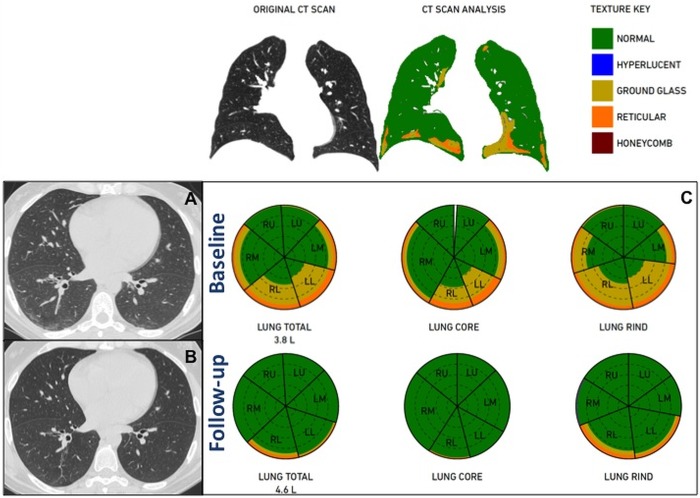
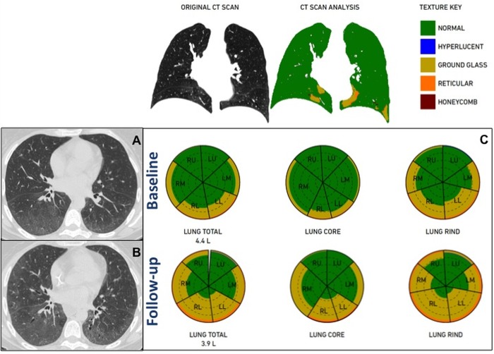
We retrospectively selected 35 consecutive patients with SSc-ILD with complete pulmonary functional evaluation, Doppler-echocardiography, immunological tests, and chest CT scan at both baseline and follow-up after immunosuppressive therapy. CT images were analyzed by two chest radiologists for semiQA and by a computational platform for texture analysis of ILD patterns (CALIPER) for QA. Concordance between semiQA and QA was tested. Traction bronchiectasis severity was scored. Analysis of ROC curves was performed.
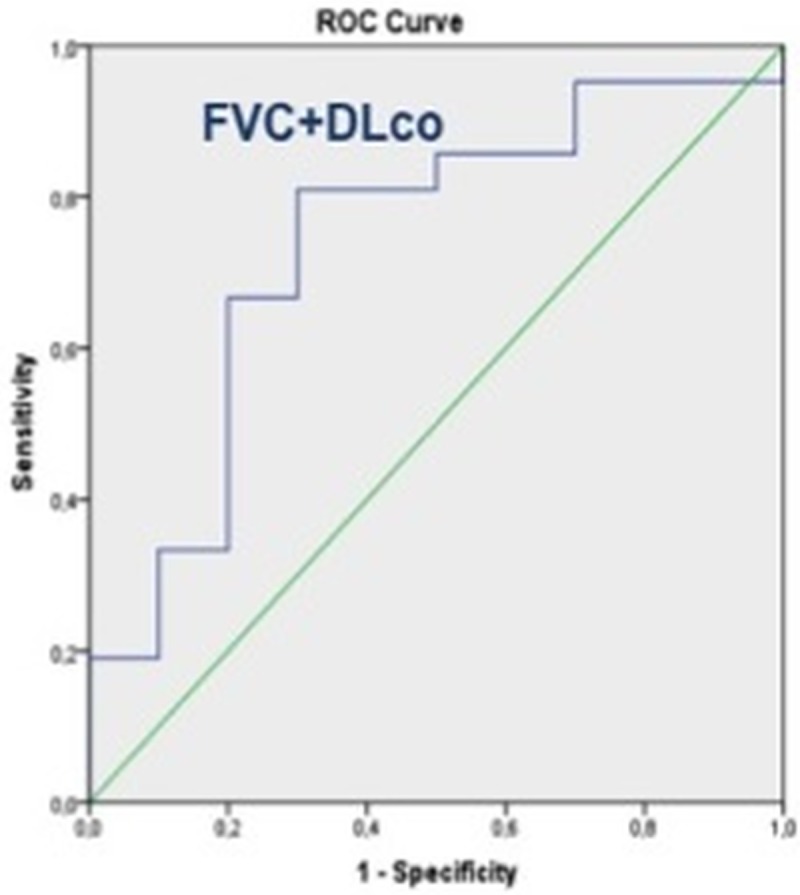
Seventy CT scans were analyzed and QA failed in 4/70 scans. Thus, the final population included 31/35 patients (51.3±12.1 years). QA had a weak-to-good concordance with semiQA (ICC reticular:0.275; ICC ground-glass:0.667) and QA correlated better than semiQA (r = -0.3 to -0.74 vs r = -0.3 to -0.4) with functional parameters. Both methods correlated with traction bronchiectases score and pulmonary artery diameter at CT. A pulmonary artery diameter ≥29mm distinguished patients with lower lung volumes and ILD extent greater than 39% (p<0.001). Changes in QA patterns during treatment were not accurate (AUC: 0.50 to 0.70; p>0.05) in predicting disease progression as assessed by functional parameters, whereas variation in total lung volume at QA accurately predicted changes in the composite functional respiratory endpoint with FVC% and DLco% (AUC = 0.74; 95%CI: 0.54 to 0.93; p = 0.03).
Pulmonary QA of CT images can objectively quantify specific patterns of ILD changes during treatment in patients with SSc-ILD. Changes in QA patterns do not correlate with functional changes, but variation in total lung volume at QA accurately predicted changes in the composite functional respiratory endpoint with FVC% and DLco%. Pulmonary artery diameter at CT reflects the interstitial involvement, identifying patients with more severe prognosis.
通过定量分析 (QA) 与胸部计算机断层扫描 (CT) 扫描的半定量分析 (semiQA) 比较,评估系统性硬化症相关间质性肺病 (SSc-ILD) 及其治疗过程中的变化。评估 QA 在预测功能变化方面的预后价值。
我们回顾性选择了 35 例 SSc-ILD 患者,这些患者均具有完整的肺功能评估、多普勒超声心动图、免疫试验和基线及免疫抑制治疗后随访的胸部 CT 扫描。两名胸部放射科医生对 CT 图像进行 semiQA 分析,并用计算平台(CALIPER)对ILD 模式进行纹理分析(QA)。测试 semiQA 和 QA 之间的一致性。评分牵引性支气管扩张的严重程度。进行 ROC 曲线分析。
分析了 70 次 CT 扫描,QA 在 4/70 次扫描中失败。因此,最终纳入 31/35 名患者(51.3±12.1 岁)。QA 与 semiQA 具有弱到良好的一致性(网状 ICC:0.275;磨玻璃 ICC:0.667),QA 与功能参数的相关性优于 semiQA(r=-0.3 到-0.74 与 r=-0.3 到-0.4)。两种方法均与 CT 上的牵引性支气管扩张评分和肺动脉直径相关。肺动脉直径≥29mm 可区分肺容积较低和 ILD 范围大于 39%的患者(p<0.001)。治疗过程中 QA 模式的变化不能准确预测功能参数评估的疾病进展(AUC:0.50 至 0.70;p>0.05),而 QA 上总肺容量的变化可准确预测 FVC%和 DLco%的复合功能呼吸终点的变化(AUC=0.74;95%CI:0.54 至 0.93;p=0.03)。
CT 图像的肺部 QA 可以客观地量化 SSc-ILD 患者治疗过程中特定的 ILD 变化模式。QA 模式的变化与功能变化不相关,但 QA 上总肺容量的变化可准确预测 FVC%和 DLco%的复合功能呼吸终点的变化。CT 上的肺动脉直径反映了间质受累情况,可识别预后更严重的患者。