Human Systems Neuroscience Laboratory, Boston University, 635 Commonwealth Ave., Room 401D, Boston, MA, 02215, USA.
Program in Neuroscience, Boston University, Boston, MA, 02215, USA.
Acta Neuropathol Commun. 2019 Mar 13;7(1):40. doi: 10.1186/s40478-019-0684-8.
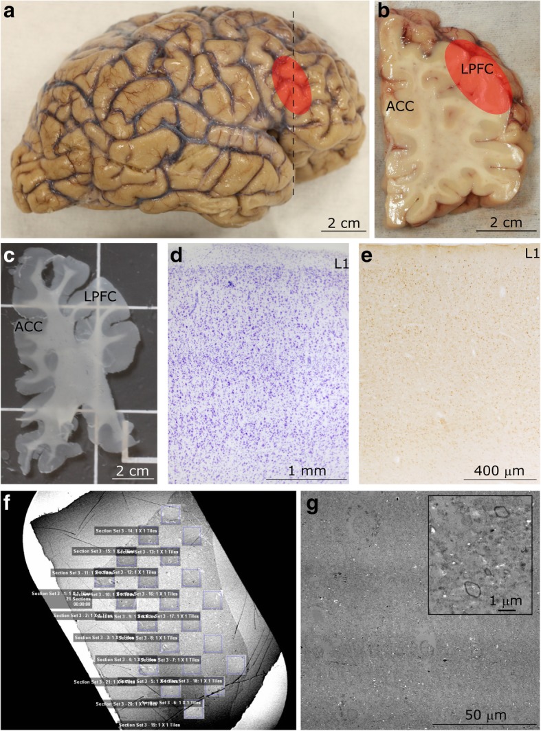
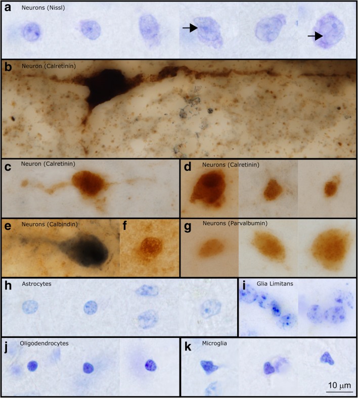
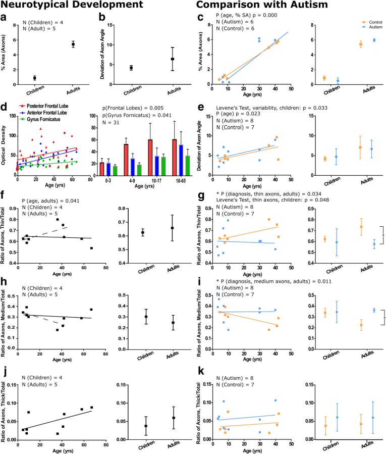
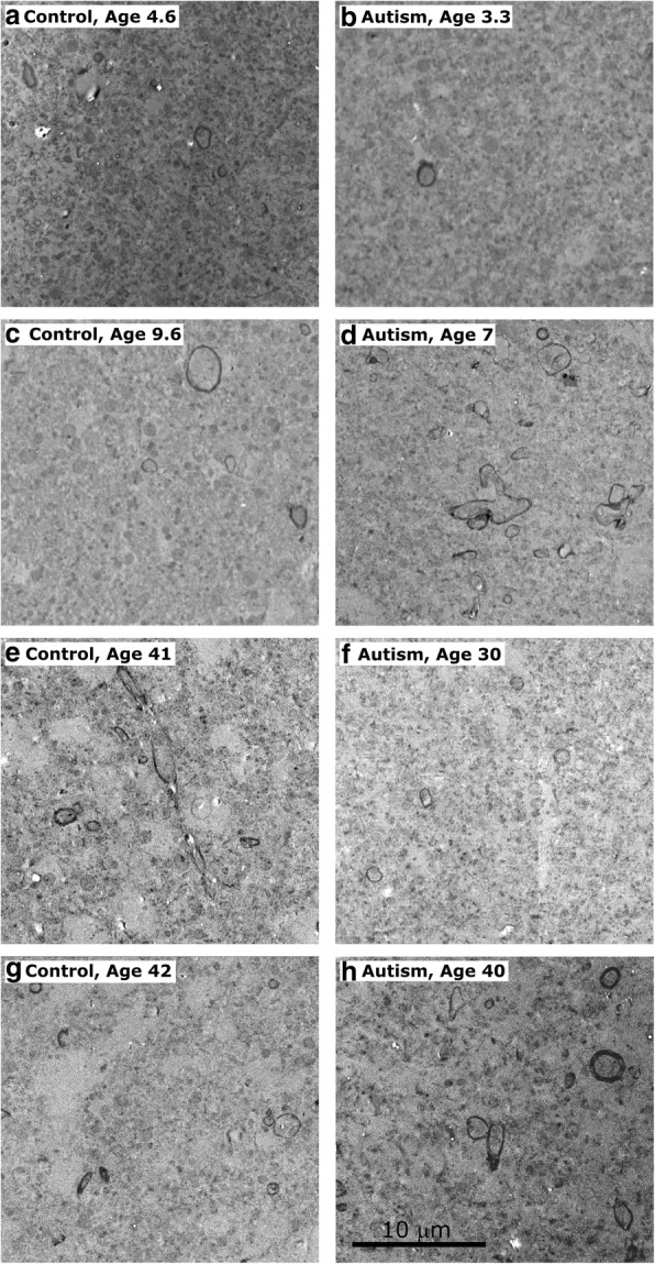
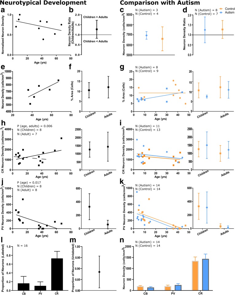
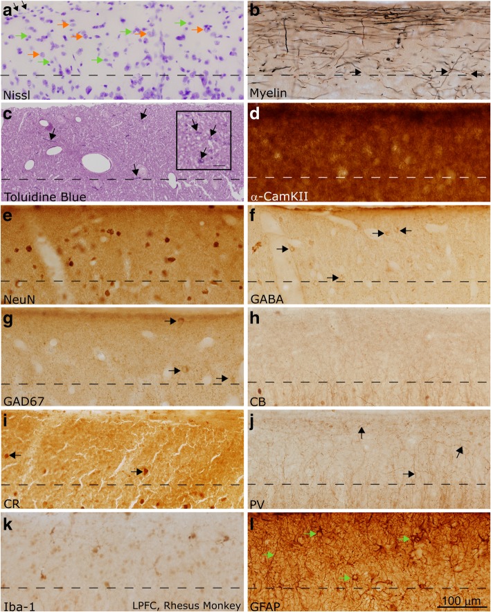
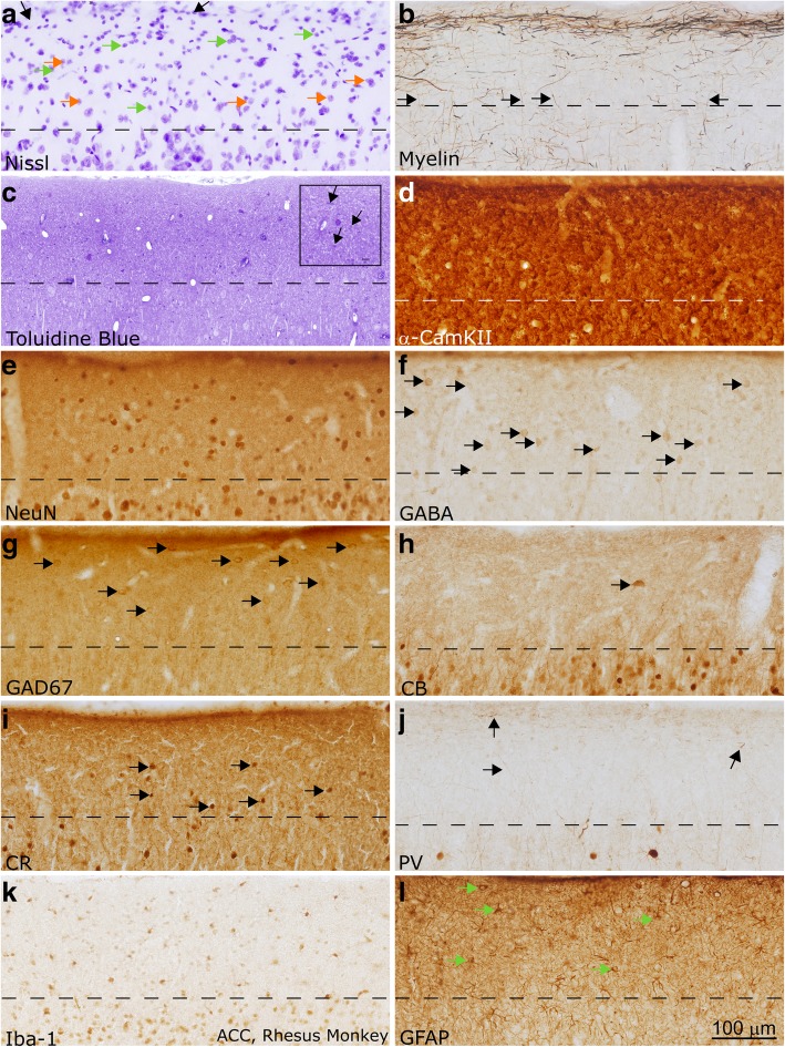
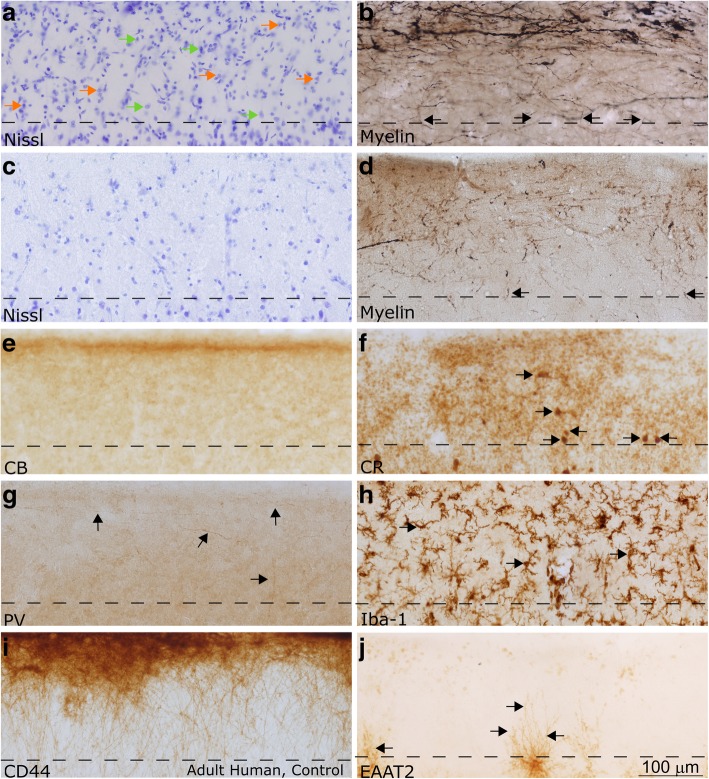
Autism is a neurodevelopmental connectivity disorder characterized by cortical network disorganization and imbalance in excitation/inhibition. However, little is known about the development of autism pathology and the disruption of laminar-specific excitatory and inhibitory cortical circuits. To begin to address these issues, we examined layer 1 of the lateral prefrontal cortex (LPFC), an area with prolonged development and maturation that is affected in autism. We focused on layer 1 because it contains a distinctive, diverse population of interneurons and glia, receives input from feedback and neuromodulatory pathways, and plays a critical role in the development, maturation, and function of the cortex. We used unbiased quantitative methods at high resolution to study the morphology, neurochemistry, distribution, and density of neurons and myelinated axons in post-mortem brain tissue from children and adults with and without autism. We cross-validated our findings through comparisons with neighboring anterior cingulate cortices and optimally-fixed non-human primate tissue. In neurotypical controls we found an increase in the density of myelinated axons from childhood to adulthood. Neuron density overall declined with age, paralleled by decreased density of inhibitory interneurons labeled by calretinin (CR), calbindin (CB), and parvalbumin (PV). Importantly, we found PV neurons in layer 1 of typically developing children, previously detected only perinatally. In autism there was disorganization of cortical networks within layer 1: children with autism had increased variability in the trajectories and thickness of myelinated axons in layer 1, while adults with autism had a reduction in the relative proportion of thin axons. Neurotypical postnatal changes in layer 1 of LPFC likely underlie refinement of cortical activity during maturation of cortical networks involved in cognition. Our findings suggest that disruption of the maturation of feedback pathways, rather than interneurons in layer 1, has a key role in the development of imbalance between excitation and inhibition in autism.
自闭症是一种神经发育性连接障碍,其特征为皮质网络组织紊乱和兴奋/抑制失衡。然而,对于自闭症病理的发展以及层特异性兴奋性和抑制性皮质回路的破坏,人们知之甚少。为了开始解决这些问题,我们研究了外侧前额叶皮质(LPFC)的第 1 层,这是一个发育和成熟时间较长且受自闭症影响的区域。我们之所以关注第 1 层,是因为它包含了独特而多样的中间神经元和神经胶质细胞群体,接收来自反馈和神经调质途径的输入,并且在皮质的发育、成熟和功能中起着关键作用。我们使用无偏置的定量方法在高分辨率下研究了来自自闭症和非自闭症儿童和成人死后脑组织中神经元和有髓轴突的形态、神经化学、分布和密度。我们通过与相邻的前扣带回皮质和最佳固定的非人类灵长类动物组织进行比较来验证我们的发现。在神经典型对照组中,我们发现从儿童期到成年期有髓轴突的密度增加。神经元密度总体随年龄下降,与之平行的是标记为 calretinin(CR)、calbindin(CB)和 parvalbumin(PV)的抑制性中间神经元密度降低。重要的是,我们在正常发育的儿童的第 1 层中发现了 PV 神经元,以前只在围产期检测到。在自闭症中,第 1 层的皮质网络出现了紊乱:自闭症儿童的第 1 层中髓鞘轴突的轨迹和厚度变化更大,而自闭症成人的薄轴突相对比例减少。LPFC 的第 1 层的神经典型的出生后变化可能是皮质网络成熟过程中皮质活动细化的基础。我们的发现表明,反馈途径的成熟障碍,而不是第 1 层中的中间神经元,在自闭症中兴奋与抑制失衡的发展中起着关键作用。