Wu Yue-Qin, Ju Chao-Long, Wang Bao-Juan, Wang Ruo-Gu
Department of Integration of Traditional Chinese Medicine and Western Medicine, Tianjin First Central Hospital, Tianjin 300192, P.R. China.
Anorectal Department of Traditional Chinese Medicine, Central Hospital of Tongchuan Mining Bureau, Tongchuan, Shanxi 727000, P.R. China.
Oncol Lett. 2019 Mar;17(3):3439-3445. doi: 10.3892/ol.2019.9999. Epub 2019 Jan 31.
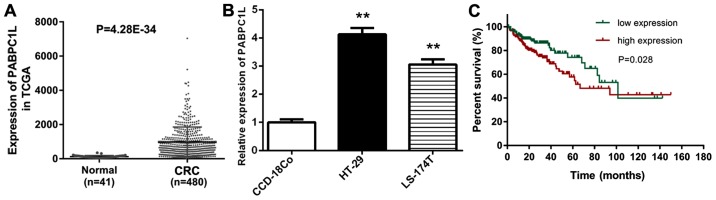
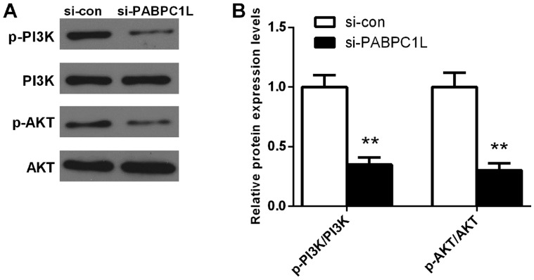
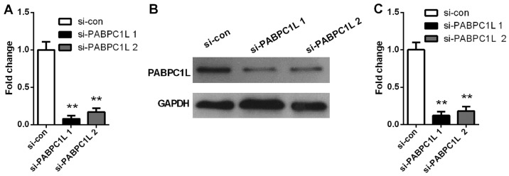
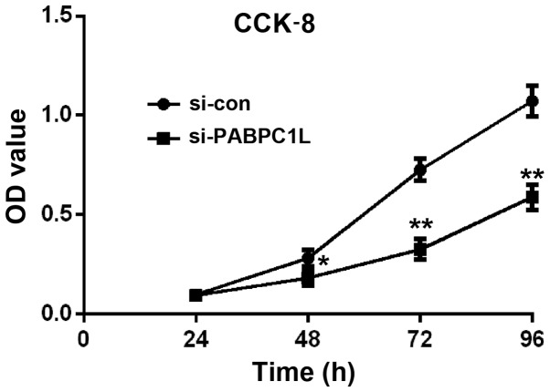
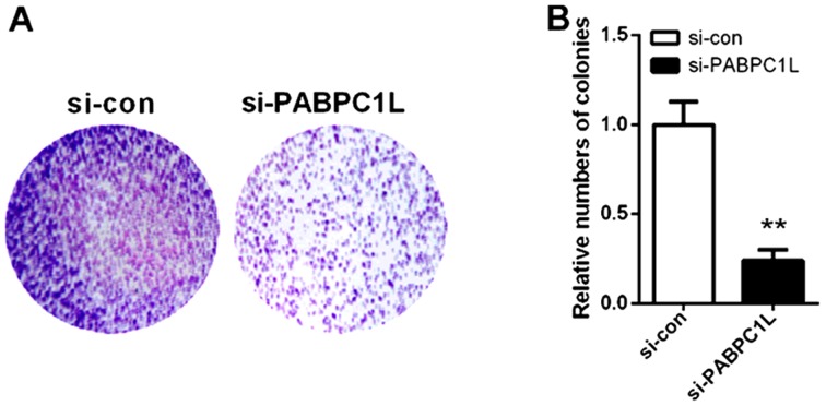
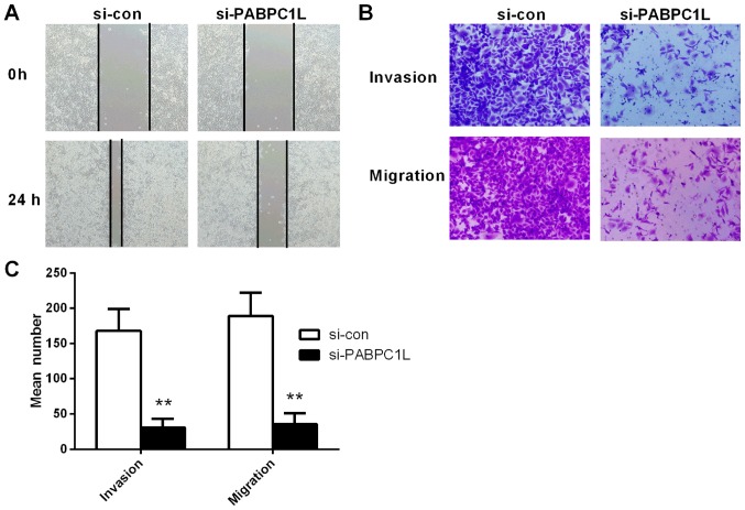
Numerous studies have demonstrated that PABPC1 participates in the process of carcinogenesis and its function is inconsistent in different types of cancers. PABPC1-like (PABPC1L) is an important paralog of PABPC1 and few studies are available on the roles of PABPC1L in colorectal cancer (CRC) development. Hence, we explored the biological function and prognostic impact of PABPC1L in CRC. The mRNA expression of PABPC1L in CRC was determined based on the data obtained from The Cancer Genome Atlas (TCGA) database. Reverse transcription-quantitative polymerase chain reaction (RT-qPCR) was utilized to determine the PABPC1L mRNA expression level in CRC HT-29 and LS-174T cell lines. Kaplan-Meier method and Cox proportional-hazards model were utilized to conduct the survival and prognosis analyses. HT-29 cells with silenced PABPC1L were constructed to explore the effect of PABPC1L on cell proliferation, invasion and migration capacities using cell counting kit-8 (CCK-8), clone formation, wound-healing and Transwell assays, respectively. To uncover the potential mechanisms of how PABPC1L influences CRC proliferation and migration, we analyzed the expression of AKT, p-AKT, PI3K, and p-PI3K in HT-29 cells using western blotting. Our results revealed that PABPC1L was overexpressed in CRC tissues compared with normal tissues based on the data obtained from TCGA database. Similarly, the mRNA expression of PABPC1L was higher in HT-29 and LS-174T cells than that in CCD-18Co cells. The expression of PABPC1L in CRC was found to be significantly related to age, pathologic stage, pathologic-node, pathologic-metastasis, and death. In univariate and multivariate analyses, pathologic-tumor and pathologic-metastasis were identified as independent prognostic factors for CRC. After PABPC1L depletion, cell proliferation rate, colony numbers, and the invasive and migratory capacity of HT-29 cells were all reduced. Western blot analysis showed that reduction of PABPC1L significantly inhibited p-AKT, and p-PI3K expression level in HT-29 cells. Collectively, our results suggested that PABPC1L is a potential novel candidate oncogene in CRC, and targeting PABPC1L may provide clinical utility in CRC.
大量研究表明,PABPC1参与致癌过程,且其在不同类型癌症中的功能并不一致。PABPC1样蛋白(PABPC1L)是PABPC1的一个重要旁系同源物,关于PABPC1L在结直肠癌(CRC)发生发展中的作用的研究较少。因此,我们探讨了PABPC1L在CRC中的生物学功能及预后影响。基于从癌症基因组图谱(TCGA)数据库获得的数据,测定了CRC中PABPC1L的mRNA表达。利用逆转录定量聚合酶链反应(RT-qPCR)来测定CRC HT-29和LS-174T细胞系中PABPC1L的mRNA表达水平。采用Kaplan-Meier法和Cox比例风险模型进行生存和预后分析。构建了PABPC1L沉默的HT-29细胞,分别使用细胞计数试剂盒-8(CCK-8)、克隆形成、伤口愈合和Transwell实验来探究PABPC1L对细胞增殖、侵袭和迁移能力的影响。为了揭示PABPC1L影响CRC增殖和迁移的潜在机制,我们使用蛋白质免疫印迹法分析了HT-29细胞中AKT、p-AKT、PI3K和p-PI3K的表达。我们的结果显示,基于TCGA数据库获得的数据,与正常组织相比,PABPC1L在CRC组织中过表达。同样,HT-29和LS-174T细胞中PABPC1L的mRNA表达高于CCD-18Co细胞。发现CRC中PABPC1L的表达与年龄、病理分期、病理淋巴结、病理转移和死亡显著相关。在单变量和多变量分析中,病理肿瘤和病理转移被确定为CRC的独立预后因素。PABPC1L缺失后,HT-29细胞的增殖率、集落数量以及侵袭和迁移能力均降低。蛋白质免疫印迹分析表明,PABPC1L的减少显著抑制了HT-29细胞中p-AKT和p-PI3K的表达水平。总的来说,我们的结果表明PABPC1L是CRC中一个潜在的新型候选癌基因,靶向PABPC1L可能在CRC中具有临床应用价值。