National Reference Centre for Parasitology, Research Institute of the McGill University Health Centre, Montreal, QC, Canada.
Program in Infectious Diseases and Immunity in Global Health, Research Institute of the McGill University Health Centre, Montreal, QC, Canada.
PLoS Negl Trop Dis. 2019 Mar 20;13(3):e0007259. doi: 10.1371/journal.pntd.0007259. eCollection 2019 Mar.
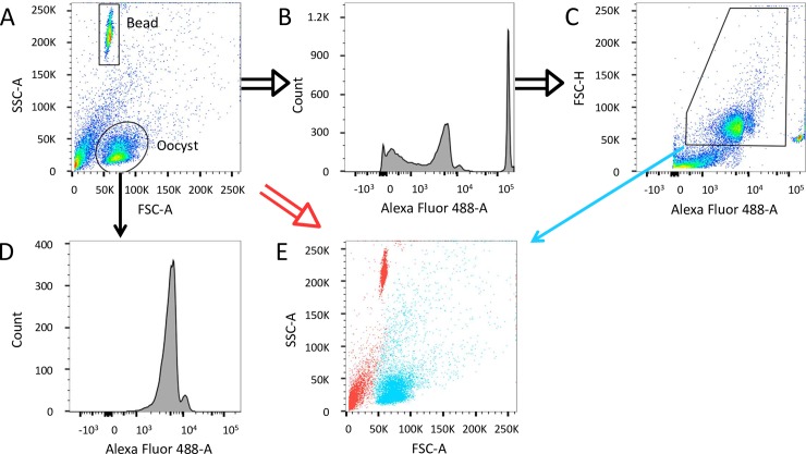
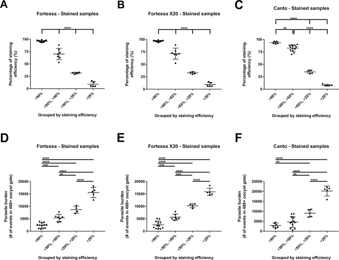
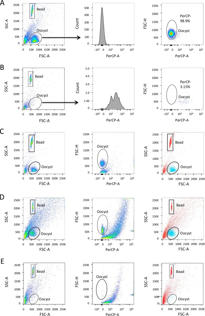
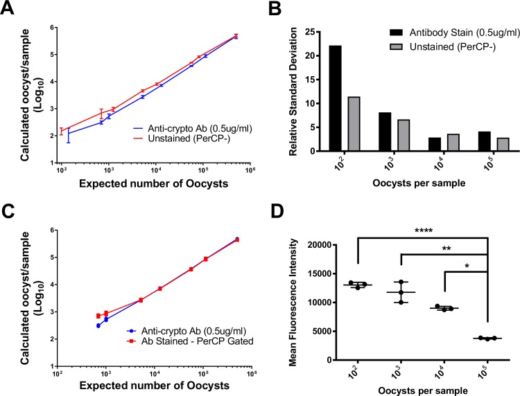
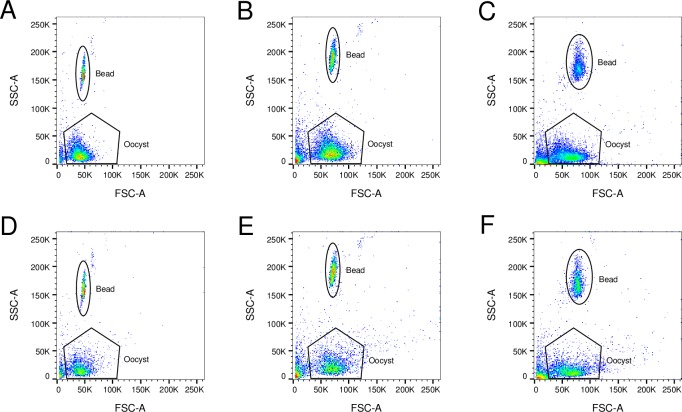
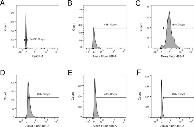
Cryptosporidiosis caused by the protozoan parasites Cryptosporidium hominis and C. parvum, threatens the lives of young children in developing countries. In veterinary medicine, C. parvum causes life-threatening diarrhea and dehydration in newborn dairy calves. Protocols to detect Cryptosporidium spp. oocysts using flow cytometry have been reported; however, these protocols use antibodies against the parasite and typically focus on detection of oocysts, not quantification. These techniques are not well-suited for studies that generate large variations in oocyst burdens because the amount of antibody required is proportional to the number of oocysts expected in samples. Also, oocysts are lost in washes in the staining protocol, reducing accuracy of oocyst counts. Moreover, these protocols require costly fluorochrome-conjugated monoclonal antibodies and are not optimal for studies involving large numbers of samples. Here we present an optimized protocol for purifying oocysts from mouse stool and intestine samples combined with a reliable method to quantify oocysts in a relatively pure population without the need for antibody staining. We used morphology (SSC-A vs FSC-A) and the innate characteristics of C. parvum oocysts compared to fecal and intestinal contaminants to develop a two-step gating strategy that can differentiate oocysts from debris. This method is a fast, reliable, and high-throughput technique to promote research projects on C. parvum infections in mice and potentially other animal hosts.
隐孢子虫病由原生动物寄生虫隐孢子虫属和小隐孢子虫引起,威胁着发展中国家幼儿的生命。在兽医领域,小隐孢子虫会导致新生奶牛犊牛发生危及生命的腹泻和脱水。已经有使用流式细胞术检测隐孢子虫属卵囊的方案报告;然而,这些方案使用针对寄生虫的抗体,通常侧重于检测卵囊,而不是定量。这些技术不适合生成卵囊负担变化较大的研究,因为所需抗体的量与预期在样本中的卵囊数量成正比。此外,在染色方案的洗涤过程中卵囊会丢失,从而降低卵囊计数的准确性。此外,这些方案需要昂贵的荧光素标记的单克隆抗体,并且不适用于涉及大量样本的研究。在这里,我们提出了一种从鼠粪便和肠组织样本中纯化卵囊的优化方案,并结合了一种无需抗体染色即可在相对纯净的群体中定量卵囊的可靠方法。我们使用形态学(SSC-A 与 FSC-A)和小隐孢子虫卵囊与粪便和肠道污染物相比的固有特征,开发了一种两步门控策略,可以将卵囊与碎片区分开来。这种方法是一种快速、可靠和高通量的技术,可以促进在小鼠和可能其他动物宿主中进行小隐孢子虫感染的研究项目。