Du Mi, Pan Wan, Duan Xiaoqi, Yang Pishan, Ge Shaohua
Shandong Provincial Key Laboratory of Oral Tissue Regeneration, Department of Periodontology, School of Stomatology, Shandong University, Jinan, PR China.
J Dent Sci. 2016 Sep;11(3):315-322. doi: 10.1016/j.jds.2016.03.009. Epub 2016 May 13.
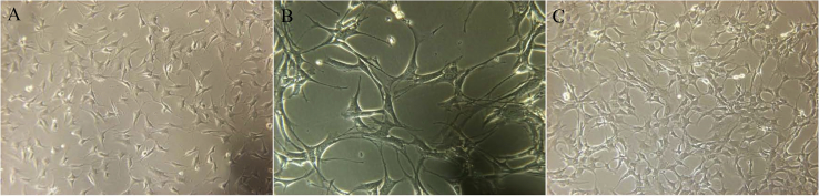
BACKGROUND/PURPOSE: The effect of aspirin on bone regeneration remains controversial. This study aimed to determine the effect of various concentrations of aspirin on cell viability, osteogenic differentiation, cell cycle, and apoptosis on ST2 cells to find an effective range of aspirin for bone regeneration induction.
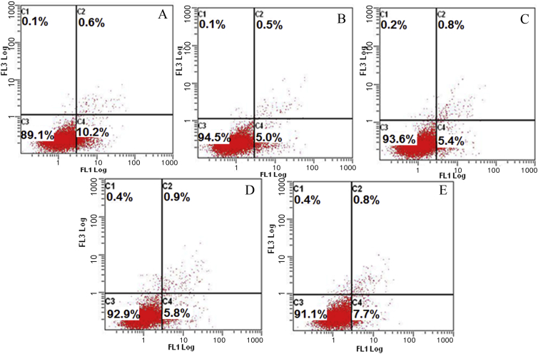
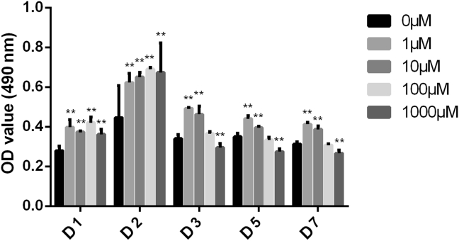
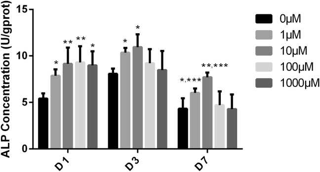
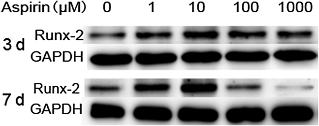
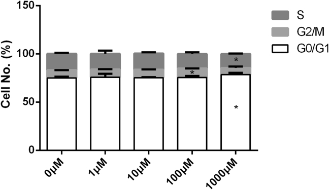
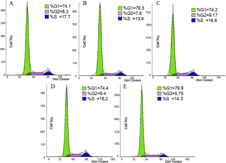
Cell viability was measured with MTT assay after being stimulated with aspirin for 1 day, 2 days, 3 days, 5 days, and 7 days. Alkaline phosphatase (ALP) activity was measured after cells were treated for 1 day, 3 days, and 7 days. Expression of runt-related transcription factor 2 (Runx-2) was evaluated using Western-blot analysis at 3 days and 7 days. Flow cytometry was used for cell cycle and apoptosis measurement after cells were treated for 48 hours.
Lower concentrations of aspirin (1μΜ and 10μM) promoted cell growth and increased ALP levels and Runx-2 expression, while higher concentrations (100μΜ and 1000μΜ) inhibited cell growth (P < 0.05), and lost their effect on ALP activity after 3 days, while even showing an inhibitory effect on the expression of Runx-2. Aspirin at a concentration of 100μM promoted cell mitosis from the S phase to the G2/M phase, and 1000μM arrested the cell cycle in the resting phase G0/G1 (P < 0.05). Parallel apoptosis/necrosis studies showed the percentage of cells in apoptosis decreased dramatically at any dose of aspirin.
A lower dosage of aspirin could promote ST2 cell growth, osteogenic differentiation, and inhibit their apoptosis which indicates that aspirin can be used as an alternative for bone regeneration.
背景/目的:阿司匹林对骨再生的影响仍存在争议。本研究旨在确定不同浓度阿司匹林对ST2细胞活力、成骨分化、细胞周期和凋亡的影响,以找到诱导骨再生的阿司匹林有效浓度范围。
用阿司匹林刺激1天、2天、3天、5天和7天后,通过MTT法测定细胞活力。细胞处理1天、3天和7天后,测定碱性磷酸酶(ALP)活性。在第3天和第7天,使用蛋白质免疫印迹分析评估矮小相关转录因子2(Runx-2)的表达。细胞处理48小时后,用流式细胞术检测细胞周期和凋亡情况。
较低浓度的阿司匹林(1μΜ和10μM)促进细胞生长,提高ALP水平和Runx-2表达,而较高浓度(100μΜ和1000μΜ)抑制细胞生长(P < 0.05),且3天后对ALP活性失去作用,甚至对Runx-2的表达有抑制作用。浓度为100μM的阿司匹林促进细胞从S期进入G2/M期进行有丝分裂,而1000μM使细胞周期停滞在静止期G0/G1(P < 0.05)。平行的凋亡/坏死研究表明,任何剂量的阿司匹林处理后,凋亡细胞的百分比均显著下降。
较低剂量的阿司匹林可促进ST2细胞生长、成骨分化并抑制其凋亡,这表明阿司匹林可作为骨再生的替代物。