Harel Mathieu, Touzet Chloe, Barthélemy Anthony, Ségard-Weisse Emilie M
Diagnostic Imaging Unit, Université de Lyon, VetAgro Sup, F-69280, Marcy l'Etoile, France.
Intensive Care Unit (SIAMU), Université de Lyon, VetAgro Sup, APCSe, F-69280, Marcy l'Etoile, France.
J Feline Med Surg. 2020 Feb;22(2):186-192. doi: 10.1177/1098612X19837336. Epub 2019 Mar 21.
The aim of this study was to report the prevalence of a honeycomb appearance of the spleen in a population of referral cats presented for ultrasound examination, and to determine the diagnostic value of this finding vs the definitive diagnosis, the splenic cytological and haematological results.
Data were obtained from the medical records (2016-2018) of cats that had an ultrasonographic honeycomb appearance of the spleen, a splenic cytological diagnosis and a complete blood count.
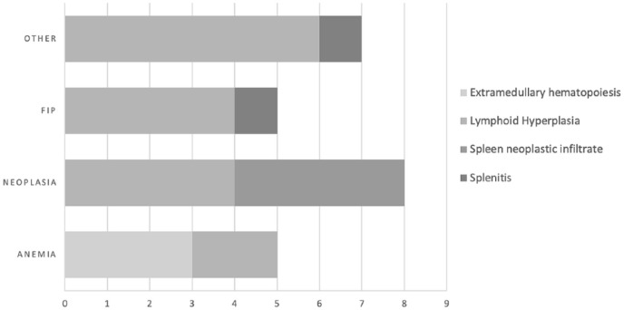
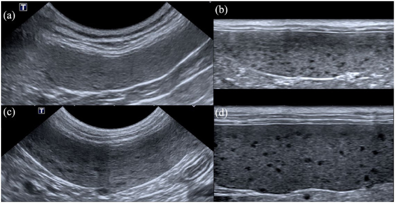
Twenty-five cats were included. Prevalence of the honeycomb pattern was 6.8%. None of the spleen was considered normal on cytology and four types of lesions were found: lymphoid hyperplasia (64%), neoplasia (16%), extramedullary haematopoiesis (12%) and splenitis (8%). A honeycomb pattern was successfully identified with a linear high-frequency probe in all cats, but only in 36% of cases with the micro-convex probe. Follow-up information was available for four cats, in which the honeycomb appearance persisted up to 105 days after the first examination; there was persistence of the honeycomb pattern in all cases. Cats with a splenic cytological diagnosis of extramedullary haematopoiesis had the lowest haemoglobin plasma concentration ( = 0.011).
Honeycomb appearance of the spleen is uncommon in cats and, in our study, was systematically associated with cytological alterations; most of the time it was benign (84%). The use of a high-frequency linear probe improves its detection rate. No epidemiological, ultrasonographic or clinical criteria allow differentiation between the different types of infiltration and fine-needle aspiration is therefore recommended.
本研究旨在报告转诊进行超声检查的猫群中脾脏蜂窝状外观的患病率,并确定这一发现相对于最终诊断、脾脏细胞学和血液学结果的诊断价值。
从2016 - 2018年有脾脏超声蜂窝状外观、脾脏细胞学诊断和全血细胞计数的猫的病历中获取数据。
纳入25只猫。蜂窝状模式的患病率为6.8%。细胞学检查未发现脾脏正常,发现了四种类型的病变:淋巴样增生(64%)、肿瘤(16%)、髓外造血(12%)和脾炎(8%)。所有猫均使用线性高频探头成功识别出蜂窝状模式,但使用微凸探头时仅在36%的病例中识别出。有4只猫有随访信息,蜂窝状外观在首次检查后持续长达105天;所有病例中蜂窝状模式均持续存在。脾脏细胞学诊断为髓外造血的猫血浆血红蛋白浓度最低( = 0.011)。
脾脏蜂窝状外观在猫中不常见,在我们的研究中,其与细胞学改变系统性相关;大多数情况下为良性(84%)。使用高频线性探头可提高其检出率。没有流行病学、超声或临床标准能够区分不同类型的浸润,因此建议进行细针穿刺抽吸。