Medical College, Yanbian University, Yanji, Jilin 133002, P.R. China.
Laboratory of Molecular Virology and Immunology, Institute of Military Veterinary Medicine, Academy of Military Medical Science, Changchun, Jilin 130122, P.R. China.
Oncol Rep. 2019 May;41(5):2818-2832. doi: 10.3892/or.2019.7077. Epub 2019 Mar 18.
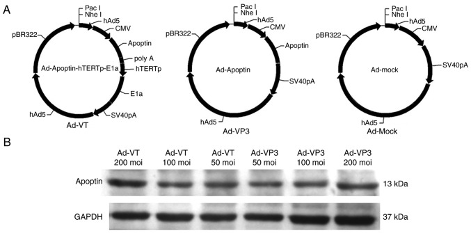
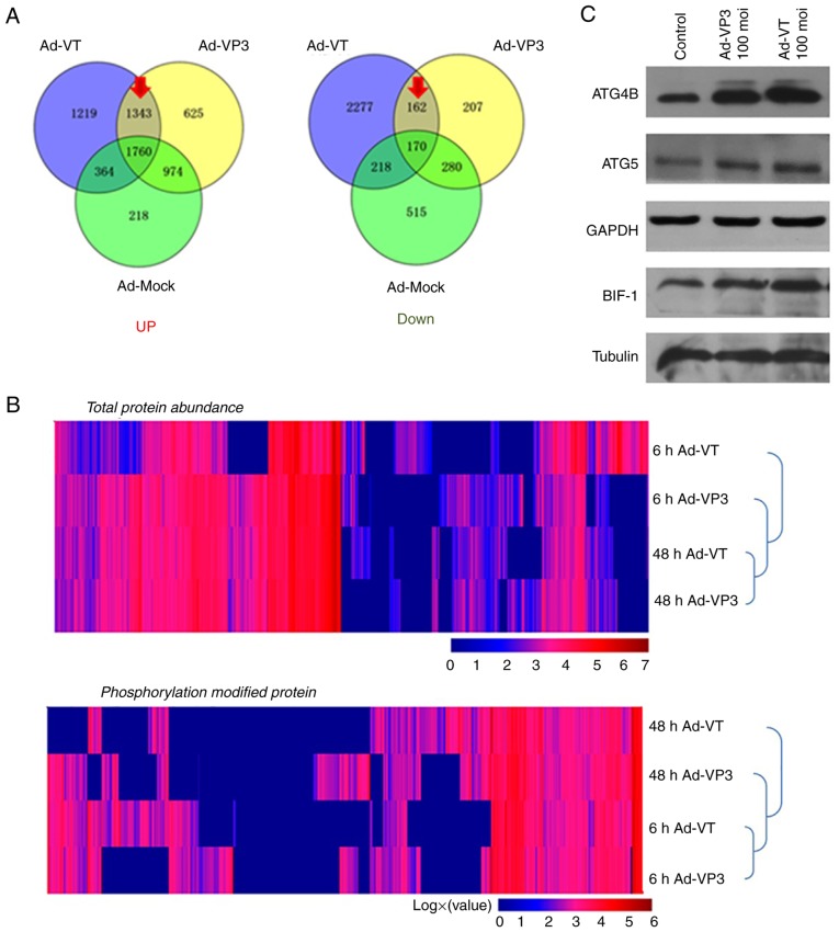
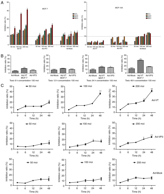
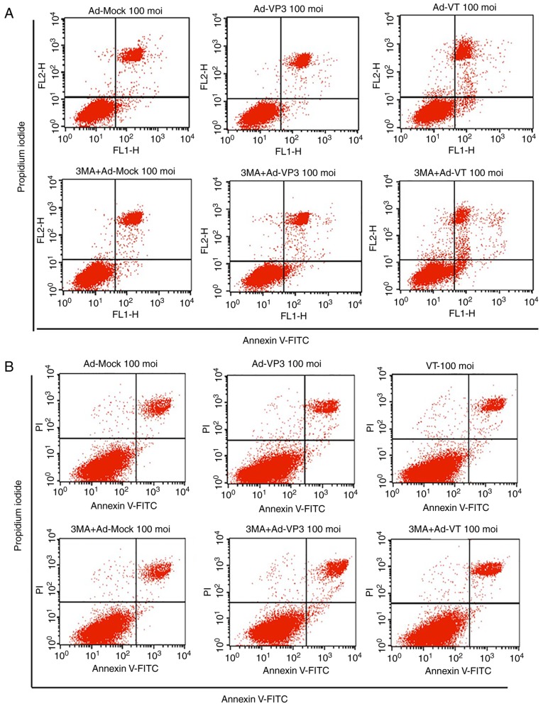
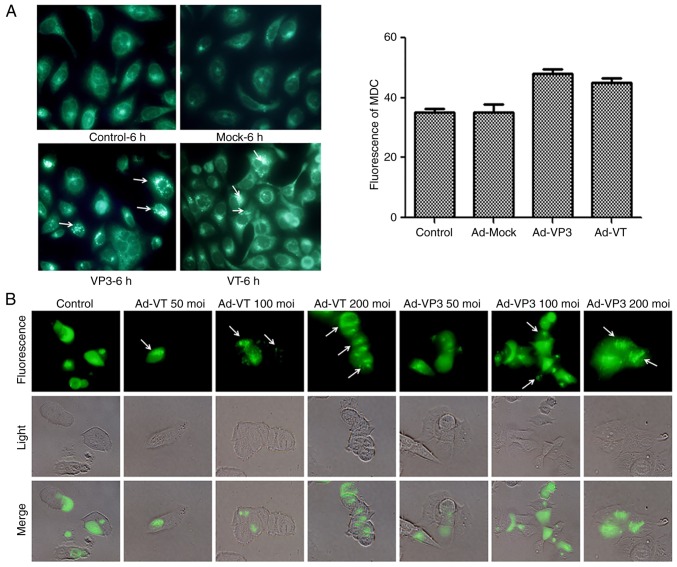
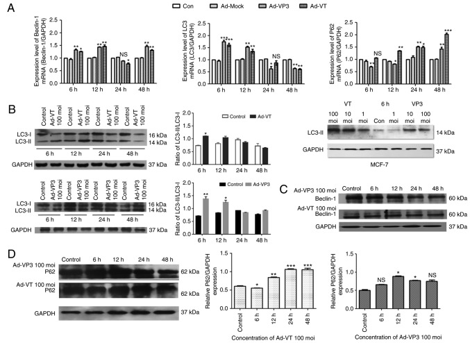
Autophagy and apoptosis both promote cell death; however, the relationship between them is subtle, and they mutually promote and antagonize each other. Apoptin can induce apoptosis of various tumor cells; however, tumor cell death is not only caused by apoptosis. Whether apoptin affects tumor cell autophagy is poorly understood. Therefore, the present study aimed to explore the potential mechanisms underlying the effects of apoptin using recombinant adenoviruses expressing apoptin. Reverse transcription‑quantitative polymerase chain reaction, immunoblotting, flow cytometry, fluorescence microscopy and proteomics analyses revealed that apoptin could induce autophagy in MCF‑7 breast cancer cells. The results also suggested that apoptin affected autophagy in a time‑ and dose‑dependent manner. During the early stage of apoptin stimulation (6 and 12 h), the expression levels of autophagy pathway‑associated proteins, including Beclin‑1, microtubule‑associated protein 1A/1B‑light chain 3, autophagy‑related 4B cysteine peptidase and autophagy‑related 5, were significantly increased, suggesting that apoptin promoted the upregulation of autophagy in MCF‑7 cells. Conversely, after 12 h of apoptin stimulation, the expression levels of apoptosis‑associated proteins were decreased, thus suggesting that apoptosis may be inhibited. Therefore, it was hypothesized that apoptin may enhance autophagy and inhibit apoptosis in MCF‑7 cells at the early stage. In conclusion, apoptin‑induced cell death may involve both autophagy and apoptosis. The induction of autophagy may inhibit apoptosis, whereas apoptosis may inhibit autophagy; however, occasionally both pathways operate at the same time and involve apoptin. This apoptin‑associated selection between tumor cell survival and death may provide a potential therapeutic strategy for breast cancer.
自噬和细胞凋亡均可促进细胞死亡;然而,它们之间的关系很微妙,相互促进又相互拮抗。凋亡素可以诱导各种肿瘤细胞的凋亡;然而,肿瘤细胞的死亡不仅是由凋亡引起的。凋亡素是否影响肿瘤细胞自噬尚不清楚。因此,本研究旨在使用表达凋亡素的重组腺病毒探讨凋亡素作用的潜在机制。逆转录-定量聚合酶链反应、免疫印迹、流式细胞术、荧光显微镜和蛋白质组学分析显示,凋亡素可诱导 MCF-7 乳腺癌细胞发生自噬。结果还表明,凋亡素以时间和剂量依赖的方式影响自噬。在凋亡素刺激的早期(6 和 12 h),自噬途径相关蛋白,包括 Beclin-1、微管相关蛋白 1A/1B-轻链 3、自噬相关蛋白 4B 半胱氨酸肽酶和自噬相关蛋白 5 的表达水平显著增加,提示凋亡素促进 MCF-7 细胞中自噬的上调。相反,在凋亡素刺激 12 h 后,凋亡相关蛋白的表达水平降低,提示凋亡可能被抑制。因此,推测凋亡素可能在早期增强 MCF-7 细胞中的自噬并抑制凋亡。总之,凋亡素诱导的细胞死亡可能涉及自噬和凋亡。自噬的诱导可能抑制凋亡,而凋亡可能抑制自噬;然而,偶尔两条途径同时作用并涉及凋亡素。这种凋亡素诱导的肿瘤细胞存活和死亡之间的选择可能为乳腺癌提供一种潜在的治疗策略。