• 文献检索
  • 文档翻译
  • 深度研究
  • 学术资讯
  • Suppr Zotero 插件Zotero 插件
  • 邀请有礼
  • 套餐&价格
  • 历史记录
应用&插件
Suppr Zotero 插件Zotero 插件浏览器插件Mac 客户端Windows 客户端微信小程序
定价
高级版会员购买积分包购买API积分包
服务
文献检索文档翻译深度研究API 文档MCP 服务
关于我们
关于 Suppr公司介绍联系我们用户协议隐私条款
关注我们

Suppr 超能文献

核心技术专利:CN118964589B侵权必究
粤ICP备2023148730 号-1Suppr @ 2026

文献检索

告别复杂PubMed语法,用中文像聊天一样搜索,搜遍4000万医学文献。AI智能推荐,让科研检索更轻松。

立即免费搜索

文件翻译

保留排版,准确专业,支持PDF/Word/PPT等文件格式,支持 12+语言互译。

免费翻译文档

深度研究

AI帮你快速写综述,25分钟生成高质量综述,智能提取关键信息,辅助科研写作。

立即免费体验

定量 CT 分析蜂巢区预测特发性肺纤维化伴明确寻常型间质性肺炎模式的死亡率:一项回顾性队列研究。

Quantitative CT analysis of honeycombing area predicts mortality in idiopathic pulmonary fibrosis with definite usual interstitial pneumonia pattern: A retrospective cohort study.

机构信息

Division of Respiratory Medicine, Department of Internal Medicine, Shiga University of Medical Science, Shiga, Japan.

Health Administration Center, Shiga University of Medical Science, Shiga, Japan.

出版信息

PLoS One. 2019 Mar 21;14(3):e0214278. doi: 10.1371/journal.pone.0214278. eCollection 2019.

DOI:10.1371/journal.pone.0214278
PMID:30897161
原文链接:https://pmc.ncbi.nlm.nih.gov/articles/PMC6428407/
Abstract

BACKGROUND

Honeycombing on high-resolution computed tomography (HRCT) images is a key finding in idiopathic pulmonary fibrosis (IPF). In IPF, honeycombing area determined by quantitative CT analysis is correlated with pulmonary function test findings. We hypothesized that quantitative CT-derived honeycombing area (HA) might predict mortality in patients with IPF.

MATERIALS AND METHODS

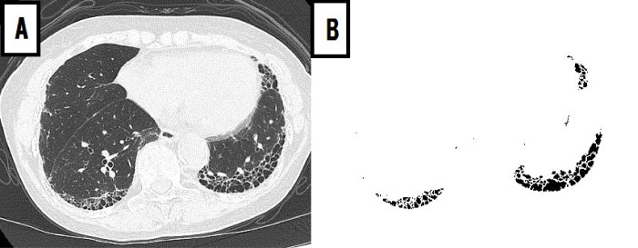
Chest HRCT images of 52 IPF patients with definite usual interstitial pneumonia (UIP) pattern were retrospectively evaluated. Mortality data up to July 31, 2016, were recorded. Using a computer-aided system, HA and percentage of HA (%HA) were measured quantitatively. Predictors of 3-year mortality were evaluated using logistic regression models.

RESULTS

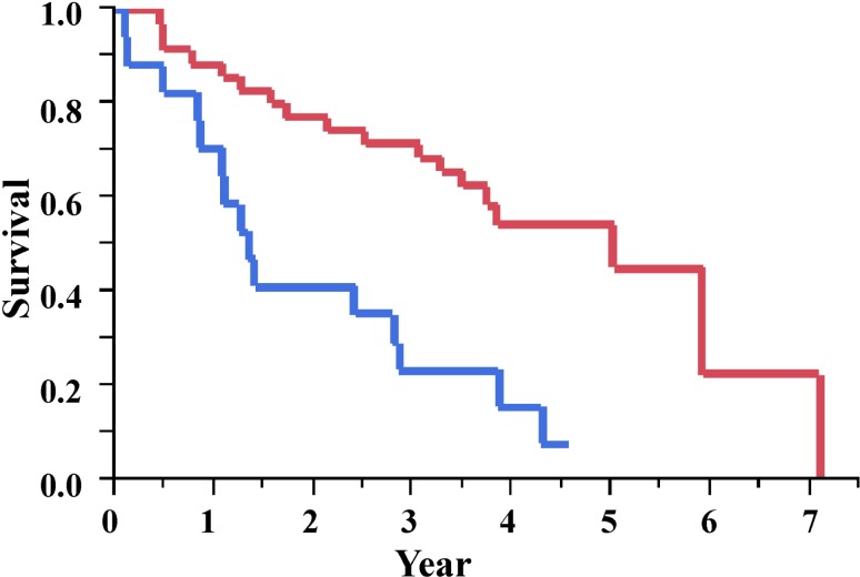
The median %HA, %predicted forced vital capacity (FVC) and composite physiologic index (CPI) were 3.8%, 83.6%, and 33.6, respectively. According to GAP (gender, age, and physiology) stage, 20, 14, and 5 patients were classified under stages I-II-III, respectively. Percentage of HA was significantly correlated with %FVC, CPI, and GAP stage (all, p < 0.001). In univariate analysis, %HA, %FVC, and CPI were statistically significant predictors of mortality. In multivariate analysis using the stepwise regression method, only %HA (odds ratio [OR], 1.27; p = 0.011) was a significant independent predictors of mortality. Patients with %HA ≥ 4.8% had significantly lower survival rates than those with lesser %HA (median survival time, 1.3 vs 5.0 years; log-rank test; p < 0.001).

CONCLUSION

Quantitative CT-derived HA might be an important and independent predictor of mortality in IPF patients with definite UIP pattern.

摘要

背景

高分辨率计算机断层扫描(HRCT)图像上的蜂巢征是特发性肺纤维化(IPF)的一个关键发现。在 IPF 中,定量 CT 分析确定的蜂巢征面积与肺功能测试结果相关。我们假设定量 CT 衍生的蜂巢征面积(HA)可能预测 IPF 患者的死亡率。

材料和方法

回顾性评估了 52 例具有明确普通间质性肺炎(UIP)模式的 IPF 患者的胸部 HRCT 图像。记录截至 2016 年 7 月 31 日的死亡率数据。使用计算机辅助系统,定量测量 HA 和 HA 百分比(%HA)。使用逻辑回归模型评估 3 年死亡率的预测因素。

结果

中位 %HA、%预计用力肺活量(FVC)和综合生理指数(CPI)分别为 3.8%、83.6%和 33.6。根据 GAP(性别、年龄和生理学)分期,20、14 和 5 例患者分别归类于 I-II-III 期。%HA 与 %FVC、CPI 和 GAP 分期均显著相关(均,p < 0.001)。在单变量分析中,%HA、%FVC 和 CPI 是死亡率的统计学显著预测因素。使用逐步回归法的多变量分析中,只有 %HA(比值比[OR],1.27;p = 0.011)是死亡率的显著独立预测因素。%HA≥4.8%的患者生存率明显低于%HA 较少的患者(中位生存时间,1.3 与 5.0 年;对数秩检验;p < 0.001)。

结论

定量 CT 衍生的 HA 可能是具有明确 UIP 模式的 IPF 患者死亡率的重要且独立的预测因素。

https://cdn.ncbi.nlm.nih.gov/pmc/blobs/1b1b/6428407/be1cb8798f96/pone.0214278.g002.jpg
https://cdn.ncbi.nlm.nih.gov/pmc/blobs/1b1b/6428407/140e3a326131/pone.0214278.g001.jpg
https://cdn.ncbi.nlm.nih.gov/pmc/blobs/1b1b/6428407/be1cb8798f96/pone.0214278.g002.jpg
https://cdn.ncbi.nlm.nih.gov/pmc/blobs/1b1b/6428407/140e3a326131/pone.0214278.g001.jpg
https://cdn.ncbi.nlm.nih.gov/pmc/blobs/1b1b/6428407/be1cb8798f96/pone.0214278.g002.jpg

相似文献

1
Quantitative CT analysis of honeycombing area predicts mortality in idiopathic pulmonary fibrosis with definite usual interstitial pneumonia pattern: A retrospective cohort study.定量 CT 分析蜂巢区预测特发性肺纤维化伴明确寻常型间质性肺炎模式的死亡率:一项回顾性队列研究。
PLoS One. 2019 Mar 21;14(3):e0214278. doi: 10.1371/journal.pone.0214278. eCollection 2019.
2
Quantitative CT analysis of honeycombing area in idiopathic pulmonary fibrosis: Correlations with pulmonary function tests.特发性肺纤维化中蜂窝状区域的定量CT分析:与肺功能测试的相关性
Eur J Radiol. 2016 Jan;85(1):125-130. doi: 10.1016/j.ejrad.2015.11.011. Epub 2015 Nov 7.
3
Clinical impact of the radiological indeterminate for usual interstitial pneumonia pattern on the diagnosis of idiopathic pulmonary fibrosis.影像学不确定的寻常型间质性肺炎模式对特发性肺纤维化诊断的临床影响。
Respir Investig. 2021 Jan;59(1):81-89. doi: 10.1016/j.resinv.2020.07.001. Epub 2020 Aug 28.
4
Normal Lung Quantification in Usual Interstitial Pneumonia Pattern: The Impact of Threshold-based Volumetric CT Analysis for the Staging of Idiopathic Pulmonary Fibrosis.普通型间质性肺炎模式下的正常肺定量分析:基于阈值的容积CT分析对特发性肺纤维化分期的影响
PLoS One. 2016 Mar 31;11(3):e0152505. doi: 10.1371/journal.pone.0152505. eCollection 2016.
5
Correlation of pulmonary function and usual interstitial pneumonia computed tomography patterns in idiopathic pulmonary fibrosis.特发性肺纤维化中肺功能与普通型间质性肺炎计算机断层扫描模式的相关性
Respir Med. 2017 Aug;129:152-157. doi: 10.1016/j.rmed.2017.06.013. Epub 2017 Jun 21.
6
Relationship of flow-volume curve pattern on pulmonary function test with clinical and radiological features in idiopathic pulmonary fibrosis.特发性肺纤维化患者肺功能检查中流量-容积曲线模式与临床及影像学特征的关系
BMC Pulm Med. 2020 Aug 12;20(1):214. doi: 10.1186/s12890-020-01254-z.
7
Comparison of clinical courses and mortality of connective tissue disease-associated interstitial pneumonias and chronic fibrosing idiopathic interstitial pneumonias.结缔组织病相关性间质性肺炎与慢性纤维化特发性间质性肺炎的临床过程和死亡率比较。
Kaohsiung J Med Sci. 2019 Jun;35(6):365-372. doi: 10.1002/kjm2.12066. Epub 2019 Mar 26.
8
Clinical impact of high-attenuation and cystic areas on computed tomography in fibrotic idiopathic interstitial pneumonias.高衰减区和囊性区域在纤维化性特发性间质性肺炎计算机断层扫描中的临床影响
BMC Pulm Med. 2015 Jul 24;15:74. doi: 10.1186/s12890-015-0069-0.
9
Possible UIP pattern on high-resolution computed tomography is associated with better survival than definite UIP in IPF patients.高分辨率计算机断层扫描上可能的 UIP 模式与特发性肺纤维化患者的明确 UIP 相比与更好的生存相关。
Respir Med. 2017 Oct;131:229-235. doi: 10.1016/j.rmed.2017.08.025. Epub 2017 Sep 12.
10
Comparison of clinical characteristics and outcomes between combined pulmonary fibrosis and emphysema associated with usual interstitial pneumonia pattern and non-usual interstitial pneumonia.合并肺纤维化和肺气肿且具有普通间质性肺炎模式与非普通间质性肺炎的临床特征及预后比较。
Sarcoidosis Vasc Diffuse Lung Dis. 2015 Jul 22;32(2):129-37.

引用本文的文献

1
Association of peripheral leukocyte telomere length with patients with rheumatoid arthritis with a focus on interstitial lung disease.外周血白细胞端粒长度与类风湿关节炎患者的关联,重点关注间质性肺疾病。
Korean J Intern Med. 2025 Jul;40(4):676-686. doi: 10.3904/kjim.2024.148. Epub 2025 Apr 1.
2
Extent of lung fibrosis is of greater prognostic importance than HRCT pattern in patients with progressive pulmonary fibrosis: data from the ILD-PRO registry.在进行性肺纤维化患者中,肺纤维化程度比高分辨率CT(HRCT)模式具有更大的预后重要性:来自ILD-PRO注册研究的数据。
Respir Res. 2025 Feb 28;26(1):73. doi: 10.1186/s12931-025-03136-6.
3

本文引用的文献

1
Computer-based quantitative computed tomography image analysis in idiopathic pulmonary fibrosis: A mini review.基于计算机的特发性肺纤维化定量计算机断层扫描图像分析:一篇综述。
Respir Investig. 2018 Jan;56(1):5-13. doi: 10.1016/j.resinv.2017.10.003. Epub 2017 Dec 7.
2
Effect of Emphysema Extent on Serial Lung Function in Patients with Idiopathic Pulmonary Fibrosis.特发性肺纤维化患者肺气肿程度对肺功能的影响。
Am J Respir Crit Care Med. 2017 Nov 1;196(9):1162-1171. doi: 10.1164/rccm.201612-2492OC.
3
Mortality prediction in idiopathic pulmonary fibrosis: evaluation of computer-based CT analysis with conventional severity measures.
The applications of CT with artificial intelligence in the prognostic model of idiopathic pulmonary fibrosis.
人工智能 CT 在特发性肺纤维化预后模型中的应用。
Ther Adv Respir Dis. 2024 Jan-Dec;18:17534666241282538. doi: 10.1177/17534666241282538.
4
Volume changes of diseased and normal areas in progressive fibrosing interstitial lung disease on inspiratory and expiratory computed tomography.进展性纤维化间质性肺疾病吸气和呼气 CT 上病变和正常区域的容积变化。
Jpn J Radiol. 2024 Aug;42(8):832-840. doi: 10.1007/s11604-024-01560-0. Epub 2024 Apr 6.
5
Radiological usual interstitial pneumonia pattern is associated with two-year mortality in patients with idiopathic pulmonary fibrosis.放射学上的普通型间质性肺炎模式与特发性肺纤维化患者的两年死亡率相关。
Heliyon. 2024 Feb 17;10(5):e26623. doi: 10.1016/j.heliyon.2024.e26623. eCollection 2024 Mar 15.
6
Quantitative analysis of high-resolution computed tomography features of idiopathic pulmonary fibrosis: a structure-function correlation study.特发性肺纤维化高分辨率计算机断层扫描特征的定量分析:一项结构-功能相关性研究。
Quant Imaging Med Surg. 2022 Jul;12(7):3655-3665. doi: 10.21037/qims-21-1232.
7
Quantitative CT in mortality prediction in pulmonary fibrosis with or without emphysema.定量CT在合并或不合并肺气肿的肺纤维化患者死亡率预测中的应用
Sarcoidosis Vasc Diffuse Lung Dis. 2021;38(3):e2021024. doi: 10.36141/svdld.v38i3.11044. Epub 2021 Sep 30.
8
Compromised Lung Volume and Hemostatic Abnormalities in COVID-19 Pneumonia: Results from an Observational Study on 510 Consecutive Patients.新型冠状病毒肺炎患者的肺容积受损和止血异常:对510例连续患者的观察性研究结果
J Clin Med. 2021 Jun 29;10(13):2894. doi: 10.3390/jcm10132894.
9
Several specific high-resolution computed tomography patterns correlate with survival in patients with idiopathic pulmonary fibrosis.几种特定的高分辨率计算机断层扫描模式与特发性肺纤维化患者的生存率相关。
J Thorac Dis. 2021 Apr;13(4):2319-2330. doi: 10.21037/jtd-20-1957.
10
In-vivo lung fibrosis staging in a bleomycin-mouse model: a new micro-CT guided densitometric approach.体内肺纤维化分期在博来霉素-小鼠模型中的应用:一种新的基于 micro-CT 的密度定量方法。
Sci Rep. 2020 Oct 30;10(1):18735. doi: 10.1038/s41598-020-71293-3.
特发性肺纤维化的死亡率预测:基于计算机的 CT 分析与传统严重程度指标的评估。
Eur Respir J. 2017 Jan 25;49(1). doi: 10.1183/13993003.01011-2016. Print 2017 Jan.
4
Automated Quantitative Computed Tomography Versus Visual Computed Tomography Scoring in Idiopathic Pulmonary Fibrosis: Validation Against Pulmonary Function.特发性肺纤维化中自动定量计算机断层扫描与视觉计算机断层扫描评分的比较:与肺功能的对照验证
J Thorac Imaging. 2016 Sep;31(5):304-11. doi: 10.1097/RTI.0000000000000220.
5
Quantitative CT analysis of honeycombing area in idiopathic pulmonary fibrosis: Correlations with pulmonary function tests.特发性肺纤维化中蜂窝状区域的定量CT分析:与肺功能测试的相关性
Eur J Radiol. 2016 Jan;85(1):125-130. doi: 10.1016/j.ejrad.2015.11.011. Epub 2015 Nov 7.
6
Visual vs Fully Automatic Histogram-Based Assessment of Idiopathic Pulmonary Fibrosis (IPF) Progression Using Sequential Multidetector Computed Tomography (MDCT).使用序列多排螺旋计算机断层扫描(MDCT)对特发性肺纤维化(IPF)进展进行基于直方图的视觉评估与全自动评估对比
PLoS One. 2015 Jun 25;10(6):e0130653. doi: 10.1371/journal.pone.0130653. eCollection 2015.
7
Validation of the GAP score in Korean patients with idiopathic pulmonary fibrosis.GAP评分在韩国特发性肺纤维化患者中的验证
Chest. 2015 Feb;147(2):430-437. doi: 10.1378/chest.14-0453.
8
High-resolution CT scoring system-based grading scale predicts the clinical outcomes in patients with idiopathic pulmonary fibrosis.高分辨率 CT 评分系统为基础的分级量表预测特发性肺纤维化患者的临床结局。
Respir Res. 2014 Jan 30;15(1):10. doi: 10.1186/1465-9921-15-10.
9
Influence of radiological emphysema on lung function test in idiopathic pulmonary fibrosis.放射性肺气肿对特发性肺纤维化肺功能检查的影响。
Respir Med. 2013 Nov;107(11):1781-8. doi: 10.1016/j.rmed.2013.08.039. Epub 2013 Sep 3.
10
Automated quantification of radiological patterns predicts survival in idiopathic pulmonary fibrosis.自动化的影像学模式定量分析可预测特发性肺纤维化患者的生存率。
Eur Respir J. 2014 Jan;43(1):204-12. doi: 10.1183/09031936.00071812. Epub 2013 Apr 5.