• 文献检索
  • 文档翻译
  • 深度研究
  • 学术资讯
  • Suppr Zotero 插件Zotero 插件
  • 邀请有礼
  • 套餐&价格
  • 历史记录
应用&插件
Suppr Zotero 插件Zotero 插件浏览器插件Mac 客户端Windows 客户端微信小程序
定价
高级版会员购买积分包购买API积分包
服务
文献检索文档翻译深度研究API 文档MCP 服务
关于我们
关于 Suppr公司介绍联系我们用户协议隐私条款
关注我们

Suppr 超能文献

核心技术专利:CN118964589B侵权必究
粤ICP备2023148730 号-1Suppr @ 2026

文献检索

告别复杂PubMed语法,用中文像聊天一样搜索,搜遍4000万医学文献。AI智能推荐,让科研检索更轻松。

立即免费搜索

文件翻译

保留排版,准确专业,支持PDF/Word/PPT等文件格式,支持 12+语言互译。

免费翻译文档

深度研究

AI帮你快速写综述,25分钟生成高质量综述,智能提取关键信息,辅助科研写作。

立即免费体验

硬膜外缩窄对大鼠脊髓脑脊液流动的影响。

Effect of extradural constriction on CSF flow in rat spinal cord.

机构信息

Faculty of Medicine and Health Sciences, Macquarie University, Technology Place, Sydney, NSW, 2109, Australia.

Elsevier Inc, John F Kennedy Boulevard, Philadelphia, PA, 19103, USA.

出版信息

Fluids Barriers CNS. 2019 Mar 26;16(1):7. doi: 10.1186/s12987-019-0127-8.

DOI:10.1186/s12987-019-0127-8
PMID:30909935
原文链接:https://pmc.ncbi.nlm.nih.gov/articles/PMC6434898/
Abstract

BACKGROUND

Fluid homeostasis in the central nervous system (CNS) is essential for normal neurological function. Cerebrospinal fluid (CSF) in the subarachnoid space and interstitial fluid circulation in the CNS parenchyma clears metabolites and neurotransmitters and removes pathogens and excess proteins. A thorough understanding of the normal physiology is required in order to understand CNS fluid disorders, including post-traumatic syringomyelia. The aim of this project was to compare fluid transport, using quantitative imaging of tracers, in the spinal cord from animals with normal and obstructed spinal subarachnoid spaces.

METHODS

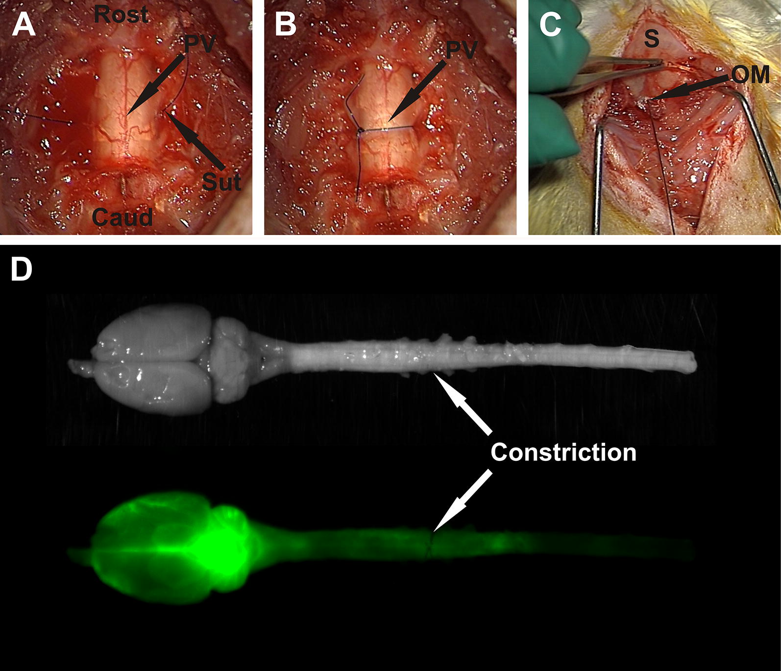
A modified extradural constriction model was used to obstruct CSF flow in the subarachnoid space at the cervicothoracic junction (C7-T1) in Sprague-Dawley rats. Alexa-Fluor 647 Ovalbumin conjugate was injected into the cisterna magna at either 1 or 6 weeks post-surgery. Macroscopic and microscopic fluorescent imaging were performed in animals sacrificed at 10 or 20 min post-injection. Tracer fluorescence intensity was compared at cervical and thoracic spinal cord levels between control and constriction animals at each post-surgery and post-injection time point. The distribution of tracer around arterioles, venules and capillaries was also compared.

RESULTS

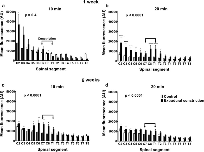
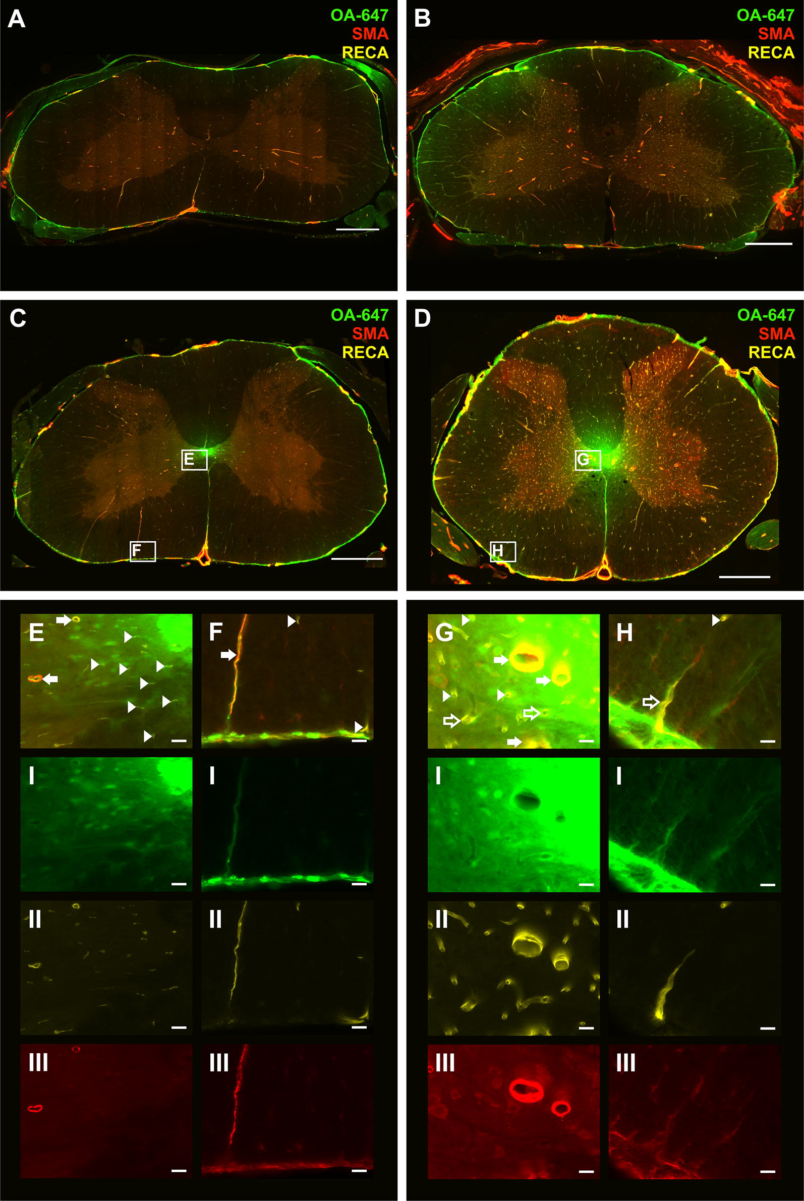
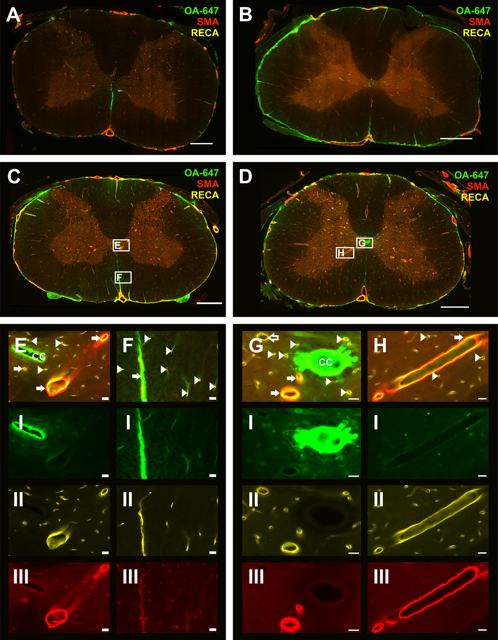
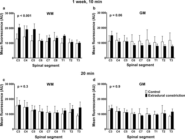
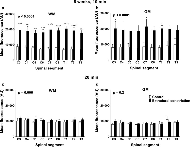
Macroscopically, the fluorescence intensity of CSF tracer was significantly greater in spinal cords from animals with a constricted subarachnoid space compared to controls, except at 1 week post-surgery and 10 min post-injection. CSF tracer fluorescence intensity from microscopic images was significantly higher in the white matter of constriction animals 1 week post surgery and 10 min post-injection. At 6 weeks post-constriction surgery, fluorescence intensity in both gray and white matter was significantly increased in animals sacrificed 10 min post-injection. At 20 min post-injection this difference was significant only in the white matter and was less prominent. CSF tracer was found predominantly in the perivascular spaces of arterioles and venules, as well as the basement membrane of capillaries, highlighting the importance of perivascular pathways in the transport of fluid and solutes in the spinal cord.

CONCLUSIONS

The presence of a subarachnoid space obstruction may lead to an increase in fluid flow within the spinal cord tissue, presenting as increased flow in the perivascular spaces of arterioles and venules, and the basement membranes of capillaries. Increased fluid retention in the spinal cord in the presence of an obstructed subarachnoid space may be a critical step in the development of post-traumatic syringomyelia.

摘要

背景

中枢神经系统(CNS)中的液体动态平衡对于正常的神经功能至关重要。蛛网膜下腔中的脑脊液(CSF)和 CNS 实质中的间质液循环可清除代谢产物和神经递质,并清除病原体和多余的蛋白质。为了理解包括创伤后脊髓空洞症在内的中枢神经系统液体紊乱,需要充分了解正常生理学。本项目旨在通过示踪剂的定量成像比较正常和阻塞性蛛网膜下腔脊髓中的液体转运。

方法

在 Sprague-Dawley 大鼠颈胸交界处(C7-T1)硬膜外使用改良的外膜缩窄模型阻塞蛛网膜下腔中的 CSF 流动。在手术后 1 或 6 周将 Alexa-Fluor 647 卵清蛋白缀合物注入枕大池。在注射后 10 或 20 分钟处死动物,进行宏观和微观荧光成像。在每个手术后和注射后时间点,比较对照和缩窄动物颈椎和胸椎脊髓水平的示踪剂荧光强度。还比较了示踪剂在小动脉、小静脉和毛细血管周围的分布。

结果

宏观上,与对照组相比,蛛网膜下腔狭窄动物的脊髓 CSF 示踪剂荧光强度明显更高,除了在手术后 1 周和注射后 10 分钟。手术后 1 周和注射后 10 分钟,狭窄动物的白质微观图像中的 CSF 示踪剂荧光强度明显更高。在缩窄手术后 6 周,在注射后 10 分钟处死的动物中,灰质和白质的荧光强度均显著增加。在注射后 20 分钟,这种差异仅在白质中显著,并且不那么明显。CSF 示踪剂主要存在于小动脉和小静脉的血管周围间隙以及毛细血管的基膜中,这突出了血管周围途径在脊髓中液体和溶质转运中的重要性。

结论

蛛网膜下腔阻塞的存在可能导致脊髓组织内的液体流量增加,表现为小动脉和小静脉的血管周围间隙以及毛细血管的基膜中的流量增加。在蛛网膜下腔阻塞的情况下,脊髓中液体的保留增加可能是创伤后脊髓空洞症发展的关键步骤。

https://cdn.ncbi.nlm.nih.gov/pmc/blobs/3c00/6434898/190e6353869c/12987_2019_127_Fig7_HTML.jpg
https://cdn.ncbi.nlm.nih.gov/pmc/blobs/3c00/6434898/fc7e47963356/12987_2019_127_Fig1_HTML.jpg
https://cdn.ncbi.nlm.nih.gov/pmc/blobs/3c00/6434898/d94cbb2f3f47/12987_2019_127_Fig2_HTML.jpg
https://cdn.ncbi.nlm.nih.gov/pmc/blobs/3c00/6434898/4ea634b70fc5/12987_2019_127_Fig3_HTML.jpg
https://cdn.ncbi.nlm.nih.gov/pmc/blobs/3c00/6434898/4558202aebcd/12987_2019_127_Fig4_HTML.jpg
https://cdn.ncbi.nlm.nih.gov/pmc/blobs/3c00/6434898/422c2f6fdeb7/12987_2019_127_Fig5_HTML.jpg
https://cdn.ncbi.nlm.nih.gov/pmc/blobs/3c00/6434898/cea5425aac14/12987_2019_127_Fig6_HTML.jpg
https://cdn.ncbi.nlm.nih.gov/pmc/blobs/3c00/6434898/190e6353869c/12987_2019_127_Fig7_HTML.jpg

相似文献

1
Effect of extradural constriction on CSF flow in rat spinal cord.硬膜外缩窄对大鼠脊髓脑脊液流动的影响。
Fluids Barriers CNS. 2019 Mar 26;16(1):7. doi: 10.1186/s12987-019-0127-8.
2
Fluid flow in an animal model of post-traumatic syringomyelia.创伤后脊髓空洞症动物模型中的流体流动
Eur Spine J. 2003 Jun;12(3):300-6. doi: 10.1007/s00586-002-0492-9. Epub 2002 Dec 6.
3
Cerebrospinal fluid flow in an animal model of noncommunicating syringomyelia.非交通性脊髓空洞症动物模型中的脑脊液流动
Neurosurgery. 1999 May;44(5):1065-75; discussion 1075-6. doi: 10.1097/00006123-199905000-00068.
4
Fluid outflow in the rat spinal cord: the role of perivascular and paravascular pathways.大鼠脊髓中的液体流出:血管周隙和血管周围途径的作用。
Fluids Barriers CNS. 2018 Apr 29;15(1):13. doi: 10.1186/s12987-018-0098-1.
5
A Poroelastic Fluid/Structure-Interaction Model of Cerebrospinal Fluid Dynamics in the Cord With Syringomyelia and Adjacent Subarachnoid-Space Stenosis.伴有脊髓空洞症和相邻蛛网膜下腔狭窄的脊髓中脑脊液动力学的多孔弹性流体/结构相互作用模型
J Biomech Eng. 2017 Jan 1;139(1). doi: 10.1115/1.4034657.
6
Altered subarachnoid space compliance and fluid flow in an animal model of posttraumatic syringomyelia.创伤后脊髓空洞症动物模型中蛛网膜下腔顺应性和脑脊液流动的改变
Spine (Phila Pa 1976). 2003 Oct 15;28(20):E413-9. doi: 10.1097/01.BRS.0000092346.83686.B9.
7
Abnormalities in spinal cord ultrastructure in a rat model of post-traumatic syringomyelia.外伤性脊髓空洞症大鼠模型中脊髓超微结构的异常。
Fluids Barriers CNS. 2020 Feb 29;17(1):11. doi: 10.1186/s12987-020-0171-4.
8
Changes in intrathoracic pressure, not arterial pulsations, exert the greatest effect on tracer influx in the spinal cord.胸内压的变化而不是动脉搏动对脊髓示踪剂流入的影响最大。
Fluids Barriers CNS. 2022 Feb 8;19(1):14. doi: 10.1186/s12987-022-00310-6.
9
Evidence for rapid fluid flow from the subarachnoid space into the spinal cord central canal in the rat.大鼠蛛网膜下腔向脊髓中央管快速液体流动的证据。
Brain Res. 1996 Jan 29;707(2):155-64. doi: 10.1016/0006-8993(95)01228-1.
10
The ultrastructure of spinal cord perivascular spaces: Implications for the circulation of cerebrospinal fluid.脊髓血管周围腔的超微结构:对脑脊液循环的影响。
Sci Rep. 2017 Oct 10;7(1):12924. doi: 10.1038/s41598-017-13455-4.

引用本文的文献

1
Cerebrospinal fluid pressure dynamics as a biomechanical marker for quantification of spinal cord compression: Conceptual framework and systematic review of clinical trials.脑脊液压力动力学作为量化脊髓压迫的生物力学标志物:概念框架与临床试验的系统评价
Brain Spine. 2025 Feb 12;5:104211. doi: 10.1016/j.bas.2025.104211. eCollection 2025.
2
Holocord syringomyelia caused by tethered cord syndrome: case report and literature review.Holocord 脊髓空洞症由脊髓栓系综合征引起:病例报告和文献复习。
BMC Neurol. 2024 Nov 11;24(1):439. doi: 10.1186/s12883-024-03951-2.
3
The Physiological Occlusion of the Central Canal May Be a Prerequisite for Syringomyelia Formation.

本文引用的文献

1
Fluid outflow in the rat spinal cord: the role of perivascular and paravascular pathways.大鼠脊髓中的液体流出:血管周隙和血管周围途径的作用。
Fluids Barriers CNS. 2018 Apr 29;15(1):13. doi: 10.1186/s12987-018-0098-1.
2
The role of brain barriers in fluid movement in the CNS: is there a 'glymphatic' system?脑屏障在中枢神经系统液体运动中的作用:是否存在“类淋巴系统”?
Acta Neuropathol. 2018 Mar;135(3):387-407. doi: 10.1007/s00401-018-1812-4. Epub 2018 Feb 10.
3
Molecular characterization of perivascular drainage pathways in the murine brain.
中央管的生理性闭塞可能是脊髓空洞症形成的一个先决条件。
Neurospine. 2023 Dec;20(4):1346-1357. doi: 10.14245/ns.2346834.417. Epub 2023 Dec 31.
4
A novel method to quantify perivascular space enlargement near the syrinx in a rodent model of post-traumatic syringomyelia.一种新型方法定量评估创伤性脊髓空洞症模型中靠近空洞处的血管周围间隙扩大。
Sci Rep. 2023 Sep 12;13(1):15043. doi: 10.1038/s41598-023-42275-y.
5
Aquaporin-4 expression and modulation in a rat model of post-traumatic syringomyelia.水通道蛋白-4 在创伤后脊髓空洞症大鼠模型中的表达和调节。
Sci Rep. 2023 Jun 14;13(1):9662. doi: 10.1038/s41598-023-36538-x.
6
Suppression of TGFβR-Smad3 pathway alleviates the syrinx induced by syringomyelia.抑制转化生长因子β受体- Smad3信号通路可减轻脊髓空洞症所致的空洞。
Cell Biosci. 2023 May 29;13(1):98. doi: 10.1186/s13578-023-01048-w.
7
Characterising spinal cerebrospinal fluid flow in the pig with phase-contrast magnetic resonance imaging.应用相位对比磁共振成像技术对猪的脊髓脑脊液流动进行特征描述。
Fluids Barriers CNS. 2023 Jan 18;20(1):5. doi: 10.1186/s12987-022-00401-4.
8
Ependymal Cilia: Physiology and Role in Hydrocephalus.室管膜纤毛:生理学及在脑积水发病机制中的作用
Front Mol Neurosci. 2022 Jul 12;15:927479. doi: 10.3389/fnmol.2022.927479. eCollection 2022.
9
Targeting choroid plexus epithelium as a novel therapeutic strategy for hydrocephalus.以脉络丛上皮细胞为靶点的新型脑积水治疗策略。
J Neuroinflammation. 2022 Jun 17;19(1):156. doi: 10.1186/s12974-022-02500-3.
10
Mapping Solute Clearance From the Mouse Hippocampus Using a 3D Imaging Cryomicrotome.使用三维成像冷冻切片机绘制小鼠海马体中的溶质清除情况。
Front Neurosci. 2021 Mar 22;15:631325. doi: 10.3389/fnins.2021.631325. eCollection 2021.
鼠脑血管周围引流途径的分子特征。
J Cereb Blood Flow Metab. 2018 Apr;38(4):669-686. doi: 10.1177/0271678X17749689. Epub 2017 Dec 28.
4
Intrathecal antibody distribution in the rat brain: surface diffusion, perivascular transport and osmotic enhancement of delivery.鞘内抗体在大鼠脑内的分布:表面扩散、血管周围转运和渗透增强递送。
J Physiol. 2018 Feb 1;596(3):445-475. doi: 10.1113/JP275105. Epub 2017 Dec 18.
5
The ultrastructure of spinal cord perivascular spaces: Implications for the circulation of cerebrospinal fluid.脊髓血管周围腔的超微结构:对脑脊液循环的影响。
Sci Rep. 2017 Oct 10;7(1):12924. doi: 10.1038/s41598-017-13455-4.
6
Arterial Pulsations cannot Drive Intramural Periarterial Drainage: Significance for β Drainage.动脉搏动无法驱动壁内动脉周围引流:对β引流的意义
Front Neurosci. 2017 Aug 24;11:475. doi: 10.3389/fnins.2017.00475. eCollection 2017.
7
Interstitial solute transport in 3D reconstructed neuropil occurs by diffusion rather than bulk flow.三维重建神经突内的间质溶质转运是通过扩散而不是体流进行的。
Proc Natl Acad Sci U S A. 2017 Sep 12;114(37):9894-9899. doi: 10.1073/pnas.1706942114. Epub 2017 Aug 28.
8
Test of the 'glymphatic' hypothesis demonstrates diffusive and aquaporin-4-independent solute transport in rodent brain parenchyma.对“glymphatic”假说的测试表明,溶质在啮齿动物脑组织中的转运具有弥散性和水通道蛋白-4 独立性。
Elife. 2017 Aug 21;6:e27679. doi: 10.7554/eLife.27679.
9
Glymphatic solute transport does not require bulk flow.脑淋巴液溶质转运不依赖于体循环。
Sci Rep. 2016 Dec 8;6:38635. doi: 10.1038/srep38635.
10
Spatial model of convective solute transport in brain extracellular space does not support a "glymphatic" mechanism.脑细胞外空间对流溶质转运的空间模型不支持“类淋巴”机制。
J Gen Physiol. 2016 Dec;148(6):489-501. doi: 10.1085/jgp.201611684. Epub 2016 Nov 11.