Medical Research Center of Shandong Provincial Qianfoshan Hospital, Shandong University, Jingshi Road 16766, Jinan, Shandong 250014, China.
Medical Imaging Department of Shandong Provincial Qianfoshan Hospital, Shandong University, Jingshi Road 16766, Jinan, Shandong 250014, China.
J Immunol Res. 2019 Mar 3;2019:6587570. doi: 10.1155/2019/6587570. eCollection 2019.
PADI4 has extensive expression in many tumors. This study applied PADI4 as a tumor marker to stimulate DC- (dendritic cell-) CIK (cytokine-induced killer), an immunotherapy approach.
A PADI4 expression plasmid was transfected into EC-originating ECA-109 cells. PADI4 gene was also inserted into a prokaryotic expression vector to produce recombinant protein. Lysate from PADI4-overexpressing cells or the purified recombinant PADI4 protein was used to load DCs, and the cells were then coincubated with CIK cells. DC and CIK cell phenotypes were determined using flow cytometry. The proliferation and viability of CIK cells were analyzed using trypan blue staining. The cytotoxic effect of DC-CIK cells on cultured ECA-109 cells was determined using CCK8 assays. Tumor-bearing mice were prepared by injection of ECA-109 cells. DC-CIK cells stimulated with lysate from PADI4-overexpressing cells or the PADI4 recombinant protein were injected into the tumor-bearing mice. The tumor growth was measured with magnetic resonance imaging (MRI).
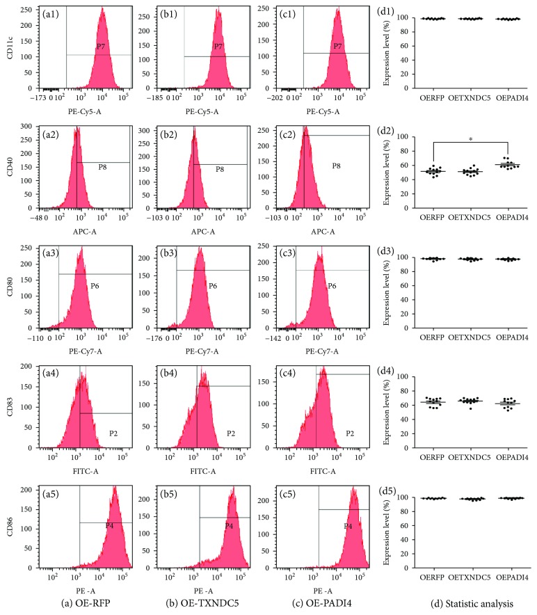
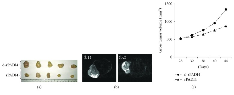
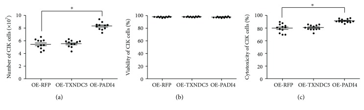
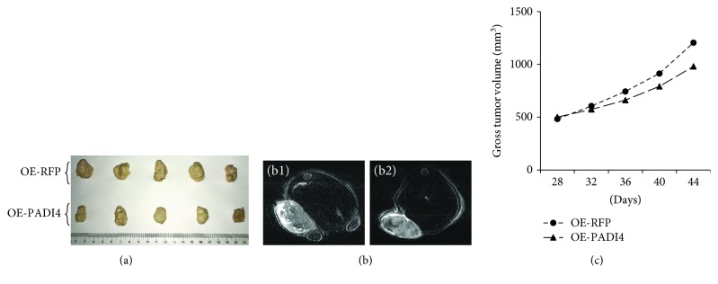
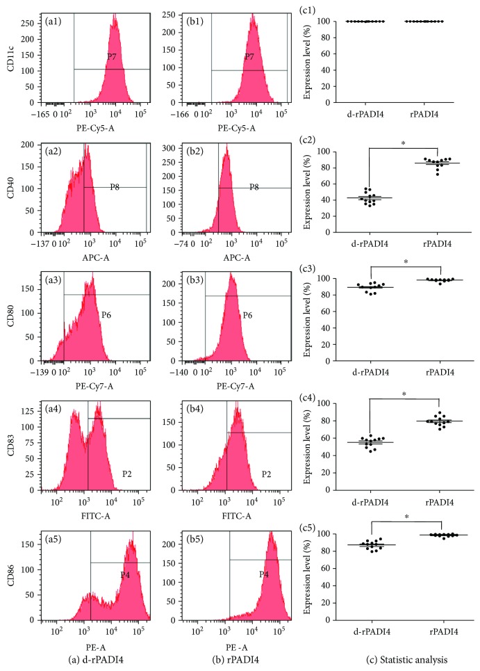
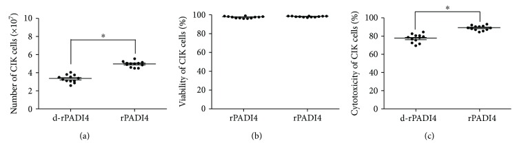
Following incubation with lysate from PADI4-overexpressing cells, the ratio of CD40 DCs increased by 17.5%. Induction of CIK cells with PADI4-stimulated DCs elevated the cell proliferation by 53.2% and the ability of CIK cells to kill ECA-109 cells by 12.1%. DC-CIK cells stimulated with lysate from PADI4-overexpressing cells suppressed tumor volume by 18.6% in the tumor-bearing mice. The recombinant PADI4 protein showed a similar effect on CIK cell proliferation and cytotoxicity as that of the lysate from PADI4-overexpressing cells. Furthermore, the recombinant protein elevated the ratio of CD40 DCs by 111.8%, CD80 DCs by 6.3%, CD83 DCs by 30.8%, and CD86 DCs by 7.8%. Induction of CIK cells with rPADI4-stimulated DCs elevated the cell proliferation by 50.3% and the ability of CIK cells to kill ECA-109 cells by 14.7% and suppressed tumor volume by 35.1% in the animal model.
This study demonstrates that stimulation of DC-CIK cells with PADI4 significantly suppressed tumor growth in tumor-bearing mice by promoting DC maturation, CIK cell proliferation, and cytotoxicity. PADI4 may be a potential tumor marker that could be used to improve the therapeutic efficiency of DC-CIK cells.
PADI4 在许多肿瘤中有广泛表达。本研究将 PADI4 作为肿瘤标志物,刺激树突状细胞-细胞因子诱导的杀伤细胞(DC-CIK)免疫治疗方法。
将 PADI4 表达质粒转染至 EC 来源的 ECA-109 细胞中。将 PADI4 基因插入原核表达载体,以产生重组蛋白。使用来自过表达 PADI4 细胞的裂解物或纯化的重组 PADI4 蛋白负载树突状细胞,然后将细胞与 CIK 细胞共培养。使用流式细胞术测定 DC 和 CIK 细胞的表型。使用台盼蓝染色分析 CIK 细胞的增殖和活力。使用 CCK8 测定法测定 DC-CIK 细胞对培养的 ECA-109 细胞的细胞毒性作用。通过注射 ECA-109 细胞制备荷瘤小鼠。将用过表达 PADI4 细胞的裂解物或 PADI4 重组蛋白刺激的 DC-CIK 细胞注入荷瘤小鼠。使用磁共振成像(MRI)测量肿瘤生长。
用来自过表达 PADI4 细胞的裂解物孵育后,CD40 DC 的比例增加了 17.5%。用 PADI4 刺激的 DC 诱导 CIK 细胞,可使细胞增殖增加 53.2%,CIK 细胞杀伤 ECA-109 细胞的能力增加 12.1%。用来自过表达 PADI4 细胞的裂解物刺激的 DC-CIK 细胞可使荷瘤小鼠的肿瘤体积抑制 18.6%。重组 PADI4 蛋白对 CIK 细胞增殖和细胞毒性的作用与来自过表达 PADI4 细胞的裂解物相似。此外,重组蛋白使 CD40 DC 的比例增加了 111.8%,CD80 DC 的比例增加了 6.3%,CD83 DC 的比例增加了 30.8%,CD86 DC 的比例增加了 7.8%。用 rPADI4 刺激的 DC 诱导 CIK 细胞,可使细胞增殖增加 50.3%,CIK 细胞杀伤 ECA-109 细胞的能力增加 14.7%,并使动物模型中的肿瘤体积抑制 35.1%。
本研究表明,用 PADI4 刺激 DC-CIK 细胞可通过促进树突状细胞成熟、CIK 细胞增殖和细胞毒性来显著抑制荷瘤小鼠的肿瘤生长。PADI4 可能是一种潜在的肿瘤标志物,可用于提高 DC-CIK 细胞的治疗效率。